

 PDF
PDF ePub
ePub Citation
 Print
Print


INTRODUCTION
Eyelid metastasis is a very rare condition that accounts for less than 1% of malignant eyelid lesions. Up to 50% of cases are derived from a breast carcinoma. Therefore, eyelid metastasis is three times more likely to occur in women than in men. However, no sex differences have been observed for metastases of other origins1. Eyelid metastases predominantly occur in adulthood, although very rare cases related to sarcoma or embryonic tumors of a neural origin can occur in children2. Cutaneous metastases from gastric adenocarcinoma are rare, accounting for only 0.8% to 1.1% of all skin metastases; they usually present as multiple subcutaneous nodules on the trunk, including Sister Mary Joseph nodule, which typically presents at the periumbilical area. Other types of cutaneous manifestations include figurate erythema, alopecia neoplastica, and erythema annulare centrifugum3. Here, we report a rare case of gastric adenocarcinoma in a patient who presented with a localized erythematous plaque on the eyelid, which is a very rare skin metastasis site.
CASE REPORT
A 75-year-old man presented with progressive left eyelid swelling that started four weeks previously. His medical history included proximal gastrectomy because of a moderately differentiated gastric adenocarcinoma five years previously. The tumor cells expressed cytoplasmic cytokeratin- 7 (CK7) but not cytokeratin-20 (CK20). The eyelid swelling improved with prednisone administration. However, leg weakness and swelling developed one week prior, and the patient was consequently transferred to the oncology department of our hospital with a suspected spinal or vertebral metastasis. Orbital magnetic resonance imaging (MRI) revealed exophthalmos and a T2-weighted high-signal-intensity lesion accompanying fatty stranding on the preseptal and septal areas. The lesion was mostly enhanced and spread to the extraconal area; the inferior rectus muscle was also thickened without any definite mass. There was no specific finding showing an acute infarct on brain MRI. The overall imaging findings were suggestive of preseptal and septal cellulitis. Therefore, teicoplanin and ceftriaxone were administered because of concern about the potential for clinical complications of orbital cellulitis including visual disturbances and central nervous system manifestations.
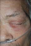
Upon physical examination at our clinic, the patient presented with localized relatively ill-demarcated erythematous swelling and indurations at the left upper and lower eyelid (Fig. 1). Histopathological examination revealed diffuse infiltration with tumor cells that formed strands or clusters between the collagen bundles throughout the upper dermis (Fig. 2A). The infiltrating tumor cells were round and polygonal and of variable size. Cytoplasm was eosinophilic, while the nuclei of the tumor cells were pleomorphic with conspicuous nucleoli with mitotic figures. Signet ring cells with intracytoplasmic mucinous vacuoles that exhibited nuclei laterally were also observed. Immunohistochemical staining showed the tumor cells expressed cytoplasmic epithelial membrane antigen (EMA) (Fig. 2B) and CK7 (Fig. 2C) but neither CK20 nor carcinoembryonic antigen (CEA). Therefore, a diagnosis of cutaneous metastasis of poorly differentiated gastric adenocarcinoma with signet ring cells was made. The patient's condition deteriorated rapidly, and he died two weeks later.
DISCUSSION
Cutaneous metastasis from an internal malignancy is uncommon, with an incidence of 5.3%; it usually arises from the breasts or lungs. Cutaneous metastasis from gastric carcinoma is rare, accounting for only 6% of all skin metastases4 and 0.8% to 1.1% of gastric cancer metastases to the skin45. Although gastric cancer is the most common type of internal malignancy in Korea, accounting for 16% of primary cancer cases, Kim et al.6 report the incidence of cutaneous metastases from gastric cancer is 0.19%. Schoenlaub et al.7 reviewed 200 cases of cutaneous metastases and report that those from gastric carcinoma were associated with a poor prognosis, with a mean survival time of 1.2 months compared to 13.8 months for breast carcinoma-derived cutaneous metastases.
Metastasis confined to the eyelid is rare, accounting for less than 1% of malignant eyelid lesions1; only three cases of eyelid metastasis have been reported8. The most common type is the nodular pattern, which accounts for up to two-thirds of all cases; it is characterized by painless subcutaneous skin-colored nodules that are often clinically mistaken for chalazionn1. The second form, which accounts for approximately one-third of cases, corresponds to the infiltrative, inflammatory, or diffuse pattern; this is characterized by painless firm swelling of the periocular skin that is usually unilateral as in the present case. This form of metastasis sometimes presents bilaterally and symmetrically, which is termed "mask-like metastasis"9. This type of metastasis can be easily misdiagnosed as orbital cellulitis or orbital contact dermatitis. Finally, the ulcerated type is due to epidermal involvement by neoplastic cells and can appear in any of the abovementioned types.
Histopathologically, metastatic gastric carcinoma exhibits aggregates of neoplastic cells in the papillary and reticular dermis that form glands, clusters, or strands embedded in an abundant fibrous stroma3. Neoplastic cells sometimes exhibit a signet ring cell morphology. The presence of signet ring cells is frequently indicative of metastatic carcinomas from the gastrointestinal tract. However, some skin tumors such as primary cutaneous signet ring cell carcinoma, liposarcoma, lymphoma, melanoma, basal cell carcinoma, and squamous cell carcinoma can also contain signet ring cells. Signet ring cell carcinoma has a greater tendency towards distant metastasis than other tumor types10. Histochemical staining is positive for Alcian blue and periodic acid Schiff stains, whereas immunohistochemical studies usually demonstrate positive immunoreactivity for CDX2, CK7, CK20, CEA, and EMA3. Hence, the present patient's history of gastric adenocarcinoma and corresponding histopathological findings confirmed the diagnosis of metastasis from gastric adenocarcinoma.
Dermatopathologists should be aware of skin metastasis from visceral malignancies because of their variable clinical appearance and presentation, which frequently delay treatment and result in failure to diagnose the condition.
For patients with malignant neoplasms, a persistent skin lesion similar to cellulitis should be suspected of being a metastasis of an internal malignancy despite the very rare occurrence of metastases at this site.

 XML Download