

 PDF
PDF ePub
ePub Citation
 Print
Print


INTRODUCTION
Alopecia areata (AA) is a common immune-mediated disease typified by multiple round areas of hair loss on the scalp. It presents as asymptomatic well-defined patches of non-scarring alopecia1. The lifetime prevalence of AA is 1.7%~2% in the United States23. The disease usually occurs in young adults, and its incidence peaks between 20 and 25 years of age2. The first disease episode typically presents before 20 years of age2. One study reports that 85.5% of Asian patients with AA present before 40 years of age4. AA in the elderly is relatively rare, thus there are few reports. A previous study indicated that late-onset AA is characterized by a marked female predominance and milder disease activity with increasing age5. The aim of our study is to better understand the demographics and clinical characteristics of AA in the elderly. In addition, we investigated the relationship between graying and the extent of AA, because the AA process preferentially targets pigmented hair, and increasing evidence indicates that the melanogenic follicular melanocytes are a principal target in AA6.
MATERIALS AND METHODS
Patient population
From January 2002 to December 2011, 1,761 patients newly diagnosed with AA were retrospectively identified who had visited the Department of Dermatology, Kyungpook National University Hospital, Daegu, Korea. Among them, 61 patients 60 years of age or older were included for analysis. However, 9 patients were lost to follow-up, 2 passed away, 10 had difficulty communicating because of impaired hearing or severe illness, and 5 refused to be interviewed. Therefore, 35 patients were finally analyzed.
Study design
We performed a retrospective study using clinical medical records and telephone interview performed by two dermatologists. The following characteristics were evaluated: sex, age of onset, duration of disease, AA severity (extent) and type, family history, past history, coexisting systemic and/or dermatologic disease, gray hair distribution at first visit, therapeutic response, and clinical course. Most data were obtained from medical records, but some were subjectively assessed by telephone interview, particularly gray hair distribution at first visit and clinical course.
For analysis of the chronological changes, the patients were divided into six age groups: 60~64, 65~69, 70~74, 75~79, 80~84, and >84 years. The types of AA were classified as basic AA (less than five patches), AA multiplex (five patches or more), alopecia totalis (entire scalp involved), and alopecia universalis (total body involved). The severity of scalp hair loss at first visit was categorized as <50%, 50%~99%, or 100% of the scalp. Hair loss on other parts of the body and nail involvement at the first consultation were evaluated in accordance with the investigative guidelines for AA by Olsen et al.7: no body hair loss (B0), some body hair loss (B1), and 100% body hair loss (B2); nail involvement was rated as none or some (N0 and N1, respectively). Treatment response with respect to baseline (i.e., first visit) was classified as better, unchanged, worse, or wax and wane. These 4 grades were also used during telephone interview to rate patients' mental and physical health status. The grade "better" was further separated into 3 subgroups according to the percentage of improved hair loss area: complete recovery (i.e., "cured"), improved area >50%, and improved area ≤50% of the alopecic area.
RESULTS
Sex ratio
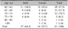
The mean age of the 61 patients was 71 years. There were 27 males (44.3%) and 34 females (55.7%) with the male-to-female ratio approximating 1:1.26 (Table 1).
Disease onset of AA and duration from onset to first visit
The age of onset of AA varied greatly. Among 61 patients, 29 (47.5%) had an age of onset from 60~64 years, which was the most common onset age group (Table 1). The duration from the recognition of initial hair loss to first hospital visit was <3 months in 16 patients (26.2%) and 3~5 months in 12 patients (19.7%).
Severity of AA at first visit
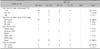
Of the 35 patients who finished the full survey, 26 (74.3%) had <50% hair loss with scalp involvement at first visit, followed by seven (20.0%) and two (5.7%) with 50%~99% and 100% hair loss with scalp involvement, respectively. Thirty-one (88.6%) patients had no hair loss on other parts of the body, and 29 (82.9%) patients also had no nail involvement (Table 2).
Clinical type of AA at first visit and ophiasis
Among the 35 AA patients, 25 (71.4%) had the basic form of AA with less than five hair loss patches; this group included two patients with ophiasis. Eight (22.9%) patients had AA multiplex, including one with ophiasis. None of the patients had alopecia totalis, and only two (5.7%) had alopecia universalis.
Past and family history of AA, and stressful events before AA onset
Three (8.6%) patients had a past history of AA. Thirty-three (94.3%) patients had no family history of AA. Fourteen (40.0%) patients reported that a stressful event occurred before AA onset.
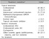
Coexisting systemic and dermatologic diseases
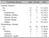
Twenty-three (65.7%) patients had coexisting systemic diseases, with hypertension being the most common. Thyroid disease (hypothyroidism) was present in only one patient. Five (14.3%) patients presented with coexisting dermatologic diseases: pruritus cutanea, urticaria, and seborrheic capitis were observed in three, one, and one, respectively (Table 3).
Gray hair in AA patients
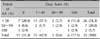
At first visit, 11 (31.4%) patients had dark hair, while 21 (68.6%) had gray hair (from 1%~100%). There was no significant association between graying and the extent of AA (p=0.679; Table 4).
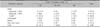
Treatment modalities
All patients except one received a combination of two or more different treatment modalities; 30 of the 34 treated patients were treated with topical steroids. Other treatment modalities included intralesional corticosteroids, minoxidil, diphenylcyclopropenone (DPCP) immunotherapy, oral medication such as a systemic steroid (low-dose continuous or minipulse), immunosuppressants (cyclosporine), antihistamines, and vitamins. Only one patient received no treatment (Table 5).
Therapeutic response and clinical course
Of the 35 patients, 22 (62.9%) showed a positive response in clinical course. Fourteen (40.0%) patients were cured, and five (14.3%) showed ≥50% improvement of the alopecic area. Four (11.4%) patients showed <50% improvement of the alopecic area. Six (17.1%) patients demonstrated unchanged clinical course, two (5.7%) worsened, and four (11.4%) showed a waxing and waning response (Table 6).
DISCUSSION
The frequency of AA ranges from 0.7%~3.8% among patients attending dermatology clinics48. AA is thought to be mediated by an autoimmune process and manifests as patchy non-scarring hair loss. AA is most commonly diagnosed in people between 20 and 25 years old2; thus, reports of AA in the elderly are rare. Previous reports of the overall population with AA show that it affects males and females equally9. However, female predominance was apparent in the present study, with a male/female ratio of 1:1.26. Wu et al.5 also reported a female predominance (male/female ratio=1:2) in late-onset AA, with first onset at age 50 years and above. However, Statistics Korea announced that overall the male/female ratio in the elderly population older than 60 years (n=7,606,903) was 1:1.34 in 2010; this rate is similar to the male/female ratio in the present study, suggesting that the sex distribution in AA in the elderly is similar to the sex distribution of the elderly in general population.
It is well documented that the prognosis of AA is proportional to the severity of the disease at onset10. The present results show that 74.3% (26/35) had an extent of hair loss <50%. This mild severity is concordant with a previous report of late-onset AA in patients aged age 50 years and above5. Yang et al.11 also report that the early-onset AA group (age of onset ≤30 years) showed a greater severity and longer duration than the late-onset group (age of onset >30 years). In the present study, 62.9% of elderly AA cases responded well to treatment. Therefore, AA in the elderly is characterized by mild clinical severity and better treatment response.
A hypothesis from a previous report on AA suggests that hair follicle melanocytes may be a primary target of immunologic attack6. In line with this hypothesis, the phenomenon of AA preferentially affecting pigmented hair but sparing graying/white hair is commonly observed5. This hypothesis can also explain the "turn white overnight" phenomenon such as 'Marie Antoinette syndrome' for the condition afflicting women and 'Thomas More syndrome' for men by selectively affecting dark hairs and leaving gray hairs12. In the present study, 31.4% (11/35) of AA patients had dark hair without graying. This rate is quite high compared to our previous report in which graying scalp hair was present in 94.2% (49/52) of AA patients with Korean ancestry at age 60~69 years13. In relation with this result, it is presumed that gray hair could be one of the reasons for the rarity of AA in the elderly. However, no significant association between gray hair and extent of AA was found in the present study.
AA is well known to be associated with atopic disease and various autoimmune diseases, particularly thyroid disease and vitiligo141415. The prevalences of atopy and thyroid diseases in AA patients are estimated to be as high as 46% and 19%, respectively58. In the present study, there were no patients with atopic dermatitis (AD) or vitiligo, and only one patient (3%) had thyroid abnormalities among 35 elderly AA patients. One-year prevalence rates of AD are much lower in the elderly than young people1617. Becerril Angeles et al.16 reported that the prevalence of AD during the senile phase (≥60 years) is 0.6%; in comparison, Rodríguez Orozco and Núñez Tapia17 reported that it is 10.1% in people aged 6~10 years. The low prevalence of older AD patients with AA observed in the present study suggests a positive relationship between the rareness of AD and old age. In contrast to the general AA population, in our study, there was only one case of thyroid disease and no cases of vitiligo among the elderly. Although such low prevalences of these diseases may be one of the features of elderly patients and the reasons for the few cases of coexisting AD and thyroid disease in the present study are unclear, the tendency of the low prevalence in the present study may be related to aging. However, a large-scale study is required to clarify this. Immunosenescence is defined as all the changes occurring in the aged immune system. Two contrasting phenomena coexist in immunosenescence: a decreasing immune response, and increased autoantibody production. The former may explain why the elderly population has decreased susceptibility to AA, like the results of our study, and the latter may demonstrate the pathogenesis of immunobullous disorders such as bullous pemphigoid, which is characterized by the production of either antibodies that react with host tissue or autoreactive immune effector T cells1819. AA and AD are immune-mediated skin diseases associated with cutaneous immune system malfunction. Therefore, the decreased ability of immune system, immunosenescence may be associated with lower incidences of both diseases. In conclusion, this study demonstrates that AA in the elderly is characterized by mild disease severity and favorable treatment response. Dark scalp hair is commonly observed in elderly AA patients. The limitations of this study include a limited review which was performed using only medical records and telephone interview and the small numbers of patients involved. The influence of aging on the pathogenesis of AA in the elderly deserves to be studied further, and larger-scale studies are required for further evaluation.
 XML Download