

 PDF
PDF ePub
ePub Citation
 Print
Print


INTRODUCTION
Atopic dermatitis (AD) is a common chronic inflammatory skin disorder that induces several symptoms including pruritus, dryness, and secondary cutaneous infection. The skin lesions of patients with AD are classified as acute or chronic according to the skin's condition and cytokine expression level in the lesional skin. Both types of atopic lesions often impair skin barrier function and exhibit decreased antimicrobial peptides expression and defective innate immune activity. Accordingly, patients with AD are more susceptible to colonization with bacteria, especially Staphylococcus aureus.
S. aureus is a well-known organism that colonizes and infects the skin in AD. The prevalence of skin colonization with S. aureus is much higher in patients with AD than that in healthy individuals: 75%~100% of patients with AD exhibit S. aureus colonization on their lesional skin, while the bacterium is isolated from only 5%~30% of individuals without AD123. Among S. aureus strains, methicillin-resistant S. aureus (MRSA) is one of the most important pathogens of community-acquired infections in many countries. Since MRSA was first reported in 1968 by Barrett et al.4, the prevalence of MRSA has increased gradually, especially in recent years. Growing evidence suggests the skin of patients with AD may be a preferred reservoir for this pathogen.
Topical antimicrobial agents are cost-effective therapies for cutaneous bacterial infections. Several approved topical agents are used to treat skin infections, many of which are usually prescribed if bacterial skin infection is suspected in AD. Among them, mupirocin and fusidic acid are the most commonly used to treat cutaneous infection5. However, the percentage of isolates resistant to these topical antibiotics has been increasing.
As antibiotic therapy plays an important role in treating bacterial skin infections in AD, the present study evaluated the antimicrobial susceptibility of S. aureus in patients with AD and determined the prevalence of resistant strains, especially MRSA. We also determined whether there are differences in the resistance rates with respect to age, because previous studies show that MRSA is more prevalent in children than in adults678. Furthermore, we determined the prevalence of resistance rates with respect to the duration of skin lesions to provide helpful information regarding the proper selection of topical antibiotics according to lesional status in AD. The results also provide information about the current prevalence of MRSA in Korea.
MATERIALS AND METHODS
The study protocol was approved by the Samsung Medical Center institutional review board (2002-10-009) and conducted in accordance with the Declaration of Helsinki. Written informed consent was obtained from all patients or their guardians.
Patients
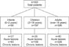
We retrospectively collected data from patients with AD who were positive for S. aureus on a skin swab performed during their first visit. A total of 583 patients including 90 infants, 187 children, and 306 adults (135 acute and 448 chronic skin lesions) who visited our dermatology outpatient clinic from July 2009 to April 2012 were recruited (Fig. 1). The exclusion criteria included were the presence of impetigo, cellulitis, or fungal infection; and the presence of other inflammatory cutaneous disorder such as psoriasis, seborrheic dermatitis, and pityriasis rosea. No patients had a history of any treatment including antibiotics in the preceding weeks. We classified AD skin lesions as acute or chronic lesions: acute lesions were defined as erythematous eczematous skin lesions with oozing and crust, while chronic lesions included erythematous to brownish papules and lichenification with xerosis.
Isolation and identification of S. aureus from AD lesions
Skin swabs for culture were taken from the lesions (i.e., oozing sites in patients with acute skin lesions or the antecubital fossa in patients with intact skin) using sterile cotton tips (BBL CultureSwab Plus; Sparks, MD, USA). Samples were inoculated onto blood agar plates, and S. aureus isolates were identified by a coagulase slide test and catalase reaction.
Antimicrobial susceptibility
The antimicrobial susceptibility of each isolate was determined by the VITEK 2 system using the AST-P601 card panel (Biomerieux, Lyon, France). The tested antibiotics included benzylpenicillin, oxacillin, gentamicin, habekacin, ciprofloxacin, telithromycin, tigecycline, erythromycin, clindamycin, quinupristin/dalfopristin, linezolid, teicoplanin, vancomycin, tetracycline, nitrofurantoin, fusidic acid, rifampicin, trimethoprim/sulfamethoxazole, and mupirocin.
Statistical analysis
Statistical significance was analyzed by the χ2 test or Fisher's exact test with a Bonferroni correction where appropriate. Logistic regression analysis was used for multivariate analysis. The level of significance was set at p<0.05. Statistical analyses were performed by using SAS version 9.4 software (SAS Institute, Cary, NC, USA).
RESULTS
Antimicrobial susceptibility patterns of S. aureus in patients with AD
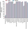
Most S. aureus isolates exhibited high susceptibility to most antimicrobial agents. The isolates exhibited less susceptibility to benzylpenicillin, erythromycin, clindamycin, and fusidic acid. In particular, isolates from chronic skin lesions showed low susceptibility to oxacillin. Among the tested antibacterial agents, S. aureus showed the highest resistance rate to benzylpenicillin followed by fusidic acid. There were no significant differences in susceptibility between acute and chronic skin lesions (Fig. 2).
Antimicrobial susceptibility of S. aureus in acute AD lesions
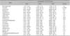
In acute cutaneous lesions, S. aureus had the lowest susceptibility to benzylpenicillin. These isolates also exhibited low susceptibility to erythromycin, clindamycin, and fusidic acid. However, all S. aureus isolates were susceptible to vancomycin, habekacin, tigecycline, quinupristin/dalfopristin, linezolid, teicoplanin, and trimethoprim/sulfamethoxazole.
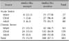
Regarding the susceptibility rates among age groups, susceptibility to oxacillin was significantly lower in the infant group (77.78% vs. 96.43% vs. 93.75% in the infant, child, and adult samples, respectively, p<0.0001). There were no significant differences in the susceptibility rates to other antimicrobial agents with respect to age group (Table 1).
Antimicrobial susceptibility of S. aureus in chronic AD lesions
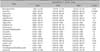
Similar to the results in patients with acute AD lesions, S. aureus from chronic lesions exhibited the lowest susceptibility to benzylpenicillin followed by fusidic acid, erythromycin, clindamycin, and oxacillin. Meanwhile, S. aureus exhibit 100% susceptibility to habekacin, linezolid, teicoplanin, vancomycin, and rifampicin.
The antimicrobial susceptibility to oxacillin was significantly lower in the infant group than the child and adult groups (p<0.0001). There were other significant differences in the antimicrobial susceptibility among the three age groups: benzylpenicillin (p=0.02), erythromycin (p=0.01), clindamycin (p=0.01), tetracycline (p=0.01), fusidic acid (p=0.04). The S. aureus isolates from the infant group were less susceptible to these antibiotics, except fusidic acid, which showed a lower susceptibility rate in the adult group (Table 2).
Isolation rates of MRSA in AD
Among the 583 S. aureus isolates, 75 (12.9%) were MRSA. The isolation rate of MRSA was significantly higher in infants with AD than that of adults or children with AD in both acute and chronic cutaneous lesions (Table 3).
Treatment response to oral antibiotics in the acute MRSA group
Among the 12 AD cases with acute skin lesions colonized with MRSA, 7 patients (4 infants, 1 child, and 2 adults) were treated with oral antimicrobial agents because of severe oozing over a large body surface area. Three patients were treated with a first-generation cephalosporin (i.e., cephradine) for two weeks, and four were treated with a second- or third-generation cephalosporin (i.e., cefuroxime and cefpodoxime). All patients taking oral cephalosporin antibiotics showed improvement of subjective symptoms and good recovery of cutaneous lesions.
DISCUSSION
MRSA (i.e., oxacillin resistant) is a major pathogen in many infectious diseases. MRSA was historically considered an important healthcare-acquired pathogen but has recently been regarded as a major cause of infection in normal populations without healthcare-associated risk factors such as long-term admission periods and intensive care unit stay. So called "community-associated MRSA" (CA-MRSA) strains are some of the most common pathogens found in skin and soft tissue infections in many countries910. One study reports the prevalence of CA-MRSA ranged from 15% to 75% among adults in 11 university-affiliated emergency departments throughout the United States11. MRSA strains account for 36%, 30%, and 23% of staphylococcal skin and soft tissue infections in North America, Latin America, and Europe, respectively12. Regarding patients with AD, several studies have investigated the incidence of MRSA isolated from AD skin lesions13141516. Hoeger14 did not identify MRSA from patients with AD in a pediatric outpatient population in 2004. However, in New Zealand, 2% of S. aureus isolates from pediatric AD cases were MRSA13. In addition, Niebuhr et al.15 found MRSA in 3% of S. aureus isolates in patients with AD. Up to 30% of S. aureus isolates from AD cases were reported to be MRSA in a Taiwanese study population in 201116. Eczematous lesions in AD are known to be a source of transmission of S. aureus. The increasing incidence rates of CA-MRSA in skin and soft tissue infections raise concerns that AD skin is a favorable reservoir for this drug-resistant organism. According to one study of the epidemiological characteristics of MRSA in Korea, 18.3% of S. aureus isolates in children with AD lesions were MRSA17.
In the present study, 12.9% (75/583) of S. aureus isolates were MRSA. MRSA was found in both acute and chronic AD lesions but more so in chronic cutaneous lesions. MRSA colonization rates are generally higher in acute skin lesions than chronic skin lesions. Our previous study of the colonization rate in AD also shows a higher S. aureus colonization rate (74%) in acute skins lesions than chronic skin lesions (38%)18. However, a large percentage of chronic AD skin lesions were colonized with MRSA (8.9% in acute lesions vs. 14.1% in chronic lesions, p>0.05). This might be explained by a history of repetitive topical antibiotics administration in chronic AD.
Interestingly, in the present study, the prevalence of MRSA was higher in the infant group regardless of lesional status. In one study, almost half of MRSA-positive children were <5 years old, and children aged between 1 month and 2 years represented just over one-third of all MRSA-positive cases19. However, it is unknown why the resistance rate is higher in infants than other age groups. Therefore, the molecular characteristics of MRSA strains by genotyping must be evaluated further to better understand about the resistance rate in infants.
The topical use of antimicrobial agents for the treatment of AD skin lesions is common and has advantages over systemic therapy with respect to cost-effectiveness and the absence of severe systemic side effects. However, the frequent use of topical antibiotics promotes the development of resistant strains. Fusidic acid is one of the most commonly used topical antibiotics in dermatology worldwide. Many studies report resistance rates against fusidic acid have increased2021222324. Accordingly, in the present study, S. aureus showed low susceptibility rates to fusidic acid in both acute and chronic lesions. In chronic AD skin lesions in particular, resistance rates to fusidic acid increased significantly with age (p=0.04). Inappropriate use of topical antibiotics leading to resistance may threaten the efficacy of systemic antibiotics for the treatment of serious S. aureus infections such as osteomyelitis and severe surgical wound infections. Therefore, the topical use of fusidic acid for empirical treatment must be restricted.
Topical mupirocin has been used since 1994 in Korea, and its use has been increasing dramatically since5. Yun et al.24 first detected mupirocin-resistant S. aureus in Korea, with a prevalence of 5%. Up to 25.3% of S. aureus isolates exhibit resistance to mupirocin in certain intensive care unit settings5. In the present study, antimicrobial susceptibility to mupirocin was relatively lower in the adult group than the infant or child group regardless of the chronicity of the lesions. The frequent and repeated use of topical mupirocin in recent years may have influenced these outcomes. Thus, awareness and research about mupirocin resistance should be bolstered for the proper long-term management of AD skin lesions. We treated 7 MRSA-positive patients with oral cephalosporin with good subjective and objective results, suggesting CA-MRSA can be controlled easily with oral cephalosporin antibiotics.
We treated 7 MRSA-positive patients with oral cephalosporin with good subjective and objective results, suggesting CA-MRSA can be controlled easily with oral cephalosporin antibiotics.
In conclusion, the prevalence of MRSA was higher in infants with AD than children and adults with AD regardless of lesional status. Furthermore, the results indicate it is rational to administer topical antibiotics susceptible to MRSA as first-line treatment for infants with AD. In addition, fusidic acid resistance was high in all age groups, and resistance rates against mupirocin tended to increase with age regardless of lesional status. This is the first study comparing the antimicrobial susceptibility rates of S. aureus isolates from AD patients of different age groups and lesional status in Korea. Thus, this study provides useful information for selecting a proper topical antimicrobial agent for patient-specific treatment according to their age and lesional status.
 XML Download