

 PDF
PDF ePub
ePub Citation
 Print
Print


INTRODUCTION
Behçet's disease (BD), first described in 1937 as a triadic complex of symptoms (oral aphthae, genital ulcers, and hypopyon uveitis), is a rare, chronic, relapsing, multisystemic idiopathic inflammatory disease. BD manifests as oral and genital ulcerations, skin lesions, and uveitis; there is also vascular, central nervous system, and gastrointestinal involvement. Vasculitis is the main histopathological finding; this is widespread, in arteries and veins of all sizes1. As to geographic distribution, BD is more prevalent along the Mediterranean coast and in Eastern Asia, with Turkey having the highest reported prevalence of between 110 and 420 per 100,000 inhabitants. In Portugal, the National Study Group for BD reported a prevalence of 2.5 per 100,000 inhabitants2. The prevalence of BD was estimated as 17 cases per 10,000 population in Kayseri, Turkey; furthermore, this previous study in Turkey reported the world's highest prevalence of BD3. Although the specific etiology remains unknown, present dogma suggests that BD is caused by unknown environmental triggering factors, ranging from infectious agents to pollution, and against a background of genetic susceptibility. The HLA-B51 gene variant is the strongest predisposition factor defined for BD so far, while other major histocompatibility complex-associated genes, such as MICA and TNF, might contribute to the disease because of their linkage disequilibrium with HLA-B512.
Since a specific laboratory test for BD is not available, its diagnosis is based exclusively on clinical assessment of patients. Several diagnostic classifications were proposed for BD, but a consensus was reached only in 1990 with the creation of the International Study Group Classification4. There have been several reports suggesting that various host genetic factors play significant roles in susceptibility to BD. Therefore, identification of the host genes responsible for susceptibility and resistance to BD should provide a significant contribution towards understanding its pathogenesis, and may lead to the development of new prophylactic and treatment strategies5.
The main approaches to treating patients with BD are symptom management, inflammation suppression at an early stage, and damage control for specific organs. Cytokines, which are crucially involved in the regulation of normal human immune responses, are important in this regard. Deregulation of cytokine expression has been shown to play a role in the pathogenesis of autoimmune diseases. Analyzing the expression of cytokines has enabled a better understanding of the pathogenesis of various diseases6.
The activation of inflammatory mechanisms in BD is considered as being governed by soluble factors, namely proinflammatory cytokines such as interleukin (IL)1, IL8, and tumor necrosis factor (TNF)-α, whose serum levels are known to increase in BD. These cytokines play a pivotal role in the activity of BD. Previous studies have shown an increase in inflammatory cytokines and chemokines, which are secreted from mononuclear phagocytes and neutrophils, in patients with BD, such as interferon-γ, TNF-α, IL1, IL2, IL6, IL8, IL12, and IL187. It was also demonstrated that the serum of patients with BD induces classical (proinflammatory) activation of human peripheral blood macrophages. Moreover, the important cytokine, IL1, also causes an increase in the serum levels of some other cytokines (such as IL8)that are reliable markers for gauging the activity of BD7.
In this study, we aimed to investigate the usability of messenger RNA (mRNA) expression of cytokine genes for following up patients with BD and also assess polymorphisms in these genes as to how they influence mRNA expression.
MATERIALS AND METHODS
Study design
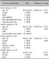
In total, 30 patients, who were referred to the Behçet Clinic of the Erciyes University Dermatology Department and diagnosed with BD according to the International Study Group Criteria for Behçet's Disease, were included in this study4. This study was approved by the Erciyes University Ethics Committee (decision number: 2008/626). The patients with BD weredivided into three groups, in order to detect any changes in the mRNA expression of IL1A, IL1B and IL2 genes in relation to pathogenesis. The first or "active" group consisted of 10 patients with active lesions, who suffered from papulopustular skin lesions, and oral and/or genital aphthae, at the time their blood was sampled; patients with oral or genital aphthous ulcers and a high level of C-reactive protein (CRP) were evaluated as active (Table 1)4. The second, "inactive," group again consisted of 10 patients but with no presently active lesions; these patients did previously have skin lesions but displayed no dermatological findings at the time their blood was sampled. A further 10 patients, who suffered posterior uveitis or panuveitis attacks at the present or any past stage of the disease, were designated as the third group, having eye involvement, the "ocular" group4. Patients with BD who also had another systemic disorder were not included in the study.
Patients in the inactive group were only receiving colchicine during the inactive phase of their disease. Patients in the active and ocular groups were receiving steroids, salazopyrin, azathioprine, methotrexate, cyclophosphamide, interferon, and anti-TNF drugs, in addition to colchicine; each patient was receiving one or more drug in combination therapy. For the active and ocular groups, patients' blood samples had been collected before they started their medical therapy.
Another group of 10 healthy subjects was included in the study as controls. Controls were evaluated by the same procedure as the cases, and were selected if they were negative for BD and any other rheumatologic or autoimmune disorder. Controls were selected from healthy subjects matched by age, sex, and ethnicity. All study subjects (patients and controls) were ethnically Turkish and from the same geographic area.
Individuals excluded from both the patient and control groups were as follows: (i) those with any metabolic or hormonal abnormality, (ii) postmenopausal women, (iii) men older than 50 years, (iv) pregnant women, (v) nursing women, and (vi) those taking medications such as estrogens, phenothiazine, cimetidine, antidepressants, sulpiride, verapamil, metoclopramide, or opiates.
There was no statistically significant difference between the patients and the controls in terms of mean age and gender. It needs be noted that, although BD is more frequent in Turkey and among the populations of countries the Silk Road used to course through (www.orpha.net), its prevalence is nevertheless quite low (1~9 per 100,000; data from www.orpha.net). This demographic impediment perforce limited the pool of patients who could be included in the study, as well as the number of controls, somewhat obscuring the level of reliability in absolutely ascertaining what role proinflammatory cytokine expression plays in the pathogenesis of BD. Nevertheless, the results can be considered as indicative where any doubt may fall on their confirmative capacity.
Determination of cytokine gene polymorphisms
Venous blood samples were taken from patients and controls for the analysis of gene polymorphisms and gene expression. DNA was isolated using the MagNA Pure LC2.0 automated isolation system (Roche, Manheim, Germany). IL1A -889(C/T) and IL1B -511(C/T) genotypes and allele frequencies were determined by restriction fragment length polymorphism (RFLP) analysis of polymerase chain reaction (PCR)-amplified DNA fragments (PCR-RFLP)7.
The IL2 -330(T/G) polymorphism was genotyped using PCR with confronting two-pair primers. PCR products were electrophoresed in 2% agarose gels containing ethidium bromide stain8.
RNA isolation, cDNA synthesis, and mRNA expression
Total RNA was isolated using TRIzol reagent (Roche) from the cells of venous blood samples. RNA concentration was determined using a NanoDrop spectrophotometer (Thermo Fisher Scientific, Waltham, MA, USA). RNA samples were stored at -80℃ until used. Total RNA (1 µg) served as a template for first-strand cDNA synthesis in a 20 µl reaction using the Transcriptor High Fidelity cDNA Synthesis Kit (Roche). The cDNA was amplified for 30 min at 55℃ and then heated for 5 min at 85℃. To ensure the fidelity of mRNA extraction and reverse transcription, all samples were normalized by PCR amplification with oligonucleotide primers specific for the constitutively expressed gene encoding β-actin. Quantitative real-time PCR (qPCR) was performed using the Roche 480 Light Cycler for the IL1A, IL1B, and IL2 genes. All samples were analyzed in duplicate. The qPCR data was analyzed using the delta cycle threshold method. The cycle threshold for each sample was calculated and relative mRNA abundance determined based on that of the β-actin control.
Statistical analysis
The relationship between BD and cytokine gene promoter polymorphisms was analyzed using a chi-square test. Logarithmic transformation (log base 2) was applied to mRNA expression data. Shapiro-Wilk's test was used to check data normality and Levene's test was used to ascertain variance homogeneity. Values are expressed as geometric means with 95% bootstrap confidence intervals obtained from 1,000 bootstrap samples. One-way analysis of variance was used to compare the difference between the groups and Tamhane's T2 test was used for pairwise comparisons. Analysis was conducted using the IBM SPSS Statistics ver. 20.0 (IBM Co., Armonk, NY, USA). A value of p<0.05 was considered statistically significant.
RESULTS
Clinical data and cytokine gene polymorphisms
Among the patients with BD included in the study within the active group, three had oral aphthous ulcers, four had oral aphthous ulcers/genital lesions, while three had papulopustular skin lesions and CRP levels higher than normal. None of the patients in the inactive group had active lesions. Six patients of the 10 included in the ocular group had posterior uveitis, whereas four were suffering from panuveitis. The clinical data of the patients included in this study are presented in Table 1.
The distribution of the IL1A -889(C/T), IL1B -511(C/T), and IL2 -330(T/G) alleles in active, inactive, and ocular patients with BD and in controls are summarized in Table 2, and the distribution of these alleles in all patients with BD compared to controls are summarized in Table 3.
A chi-square test was applied in order to compare the IL1A -889(C/T), IL1B -511(C/T), and IL2 -330(T/G) polymorphisms among the study groups, and no significant differences were recorded for the IL1A -889(C/T) and IL1B -511(C/T) polymorphisms. A higher frequency of the IL2 -330 G allele was found to be statistically significant in BD patients compared to controls (p=0.006, Table 2; p=0.011, Table 3).
mRNA expression of the IL1A, IL1B, and IL2 genes
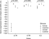
Significant differences in IL1A gene expression level were observed between the patient groups. A decreased IL1A gene expression level was found in the active patient group compared to controls (p<0.001; Table 4), whereas, an increased expression level was found in the inactive and ocular groups compared to the control group. Among the patient groups, IL1α gene expression levels were highest in the inactive group while they were recorded at remarkably low levels in the active group (Table 4). IL1B gene expression displayed significant differences between the groups; increased IL1B gene expression levels were found in the active, inactive, and ocular groups compared to controls (Table 4). IL2 mRNA expression levels also manifested no significant change in the active group in comparison with the control group, but increased in the inactive and ocular groups.
DISCUSSION
Current evidence suggests that genetic, environmental, and immunologic factors may play a role in the development of BD9,10.
We examined promoter polymorphisms of the IL1A, IL1B, and IL2 genes and their mRNA expression in patient groups with BD in the active, inactive, or ocular phase, and in a control group. No significant difference was found in the genotype distribution or allele frequency of the IL1A -889(C/T) polymorphism between patients and controls in our study. Previous studies demonstrated that the presence of the IL1A -889C allele was significantly associated with BD10,11,12, although Coskun et al.7 did not observe any such relationship.
There are studies that support a functional polymorphism in the IL1B gene possibly playing a role in BD etiology, as IL1β plays an important role in the initiation of the proinflammatory process in BD10. No significant difference was found in the genotype distribution or allele frequency of the IL1B -511(C/T) polymorphism between patients with BD and controls in our study.
In the present study, mRNA expression of the IL1A, IL1B, and IL2 genes in BD was also examined. Some studies reported elevated serum levels of the IL1β protein in patients with BD but no correlation with disease activity13,14.
Furthermore, an increased expression of synovial IL1β in BD patients was observed in previous studies13,15,16. Evereklioglu et al.17 did not detect any difference between patients with active or inactive BD and controls in respect of their serum levels of IL1β. In our study, the IL1B gene expression level was significantly increased in the active, inactive, and ocular groups compared to the control group (Fig. 1).
We also found statistically significant differences in mRNA expression level of the IL1A gene in the active, inactive, and ocular groups compared to the control group (Fig. 1); it was increased in the inactive and ocular groups compared to the controls, and decreased in the active group in comparison with the other groups (Table 4; Fig. 1). The change in the expression level of the IL1A gene in different patient groups strongly indicates that gene analysis could be useful in the prognosis and treatment of BD.
IL2 plays an important role in the pathogenesis of BD and the IL2 gene therefore attracts much attention as a candidate locus for BD. IL2 is produced and secreted by T cells and is crucial for the stimulation of T cell proliferation18. Secreted IL2 stimulates autocrine and paracrine IL2 receptor (IL2R) signaling19. Inadequate production of IL2 may cause an increase in the number of activated autoreactive cells19,20.
The importance of the IL2/IL2R system for T cell homoeostasis, at the levels of repertoire selection, generation of suppressive regulatory T cells, T cell homing, and clonal contraction via activation-induced cell death, indicates its relevance to the development of autoimmune disease20. Deficient production of IL2 may cause an increase in the number of activated autoreactive cells19,20.
We examined the IL2 -330(T/G) polymorphism and IL2 mRNA expression in this study; statistically significant increases were found in the frequencies of the IL2 -330 GG and TG genotypes in patients with BD compared to controls (Table 3). Alayli et al.11 found no significant difference between patients with BD and a control group in terms of the IL2 -330(G/T) polymorphism. However, in 2011, Shahram et al.21 reported that BD was associated with the GG genotype of the IL2 -330(T/G) polymorphism.
Furthermore, we also examined mRNA expression of the IL2 gene. In our study, we found statistically significant reduced mRNA expression in the active group compared to the control group. The results obtained in this study, increased frequency of the IL2 -330 G allele along with reduced mRNA expression of the IL2 gene in the active group, led us to believe that the IL2 gene -330 G allele may function to reduce mRNA expression of the IL2 gene, and explain the lower IL2 mRNA levels in the active group compared to the controls. Information concerning the mRNA expression of the IL2 gene in BD is not available in the literature. Other studies investigated the IL2 protein and reported an increase in serum levels of IL2 in patients with BD compared to a control group22. Significant differences in IL2 serum levels were not found between patients with BD and controls in two other studies conducted in Turkey17,23. Differences between the serum IL2 protein and IL2 mRNA expression data maybe related to regulation occurring at the transcriptional level.
BD is a complex disease, different patients will manifest different symptoms, and there is a clear geographical distribution to its prevalence. The candidate-gene approach has been useful in identifying genes affecting the susceptibility to and severity of BD. Recent scientific discoveries pertaining to the pathogenesis of BD have demonstrated the important roles of genetic and immunologic factors. BD is characterized by a divergence in the production profile of cytokines, in which several factors are required for optimal induction of T helper (Th)1 activity10,11,19. There is evidence that an imbalance in the polarization of Th1/Th2 cells in favor of Th1 cells may be the key to the pathogenic mechanism of chronic inflammatory diseases, as well as organ-specific autoimmune diseases like BD. An overproduction of proinflammatory cytokines from several cellular sources appears to be responsible for the inflammatory reaction in BD, with levels of interferon-γ, TNF-α, IL6, IL8, and IL12 being higher in patients with BD22,23.
The expression and promoter polymorphisms of genes have been determined using the same patient and control groups, so as to record how polymorphisms have had an impact on the expression of genes, because promoter sequences are potential sites of polymorphisms affecting gene expression. Promoters are involved in initiating transcription and are therefore among the many important cis-acting elements that regulate gene expression and that might harbor functionally relevant polymorphisms24.
The relatively small sample size imposes limitations on the statistical strength of this study. Admittedly, the inclusion of a few further samples may have had an effect upon the range of expression for genes reported here as, most importantly, the number of patients with BD was not sufficient for such a study. However, the results are sufficient to justify our conviction that further studies along the same lines, with larger and diversified population groups, need to be performed to test our findings. Our study reasonably demonstrates that this approach is capable of opening a diagnostic and prognostic vista for assaying the progression of BD; the decrease, at the incipient phase of BD, in the mRNA expression levels of the IL1A and IL2 genes within the active group compared to the control group, as the IL2 and IL1A mRNA expression levels increase in the inactive and ocular groups relative to the control group, certainly seems to point in that direction.
In the light of its findings, and despite its limitations, our study demonstrates that promoter polymorphisms and mRNA expression of the IL1A, IL1B, and IL2 genes indicate susceptibility to BD. We believe that the results reveal the importance of achieving a better understanding of BD and the prospects of developing therapeutic strategies for the future.


 XML Download