

 PDF
PDF ePub
ePub Citation
 Print
Print


Dear Editor:
Actinic granuloma is a rare inflammatory skin disorder that presents in chronically sun-damaged skin with flesh-colored to erythematous papules that coalesce to form centrifugally enlarging annular patterns1. However, its pathogenesis remains to be elusive. It is thought that ultraviolet radiation, heat, viral diseases or other unknown factors transform the antigenicity of elastic fibers and induce cellular immune reactions1,2.
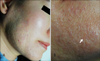
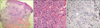
A 49-year-old Korean woman had reddish patches on the right side of her face from birth. She was diagnosed with capillary malformation about 20 years ago. She has treated the skin lesion with a pulsed dye laser (PDL) and fractional laser >10 times with 7 years. However, her skin lesion has not completely disappeared. On examination, the reddish patch had an uneven surface and firm texture (Fig. 1). These changes in the skin occurred 2 years ago. Histopathologically, there were some telangiectatic vessels and granulomatous inflammation in the superficial and mid-dermis (Fig. 2A). The dermis showed histiocytes with numerous giant cells, and some of them contained elastotic material, which was consistent with actinic granuloma (Fig. 2B, C). Because of previous repeated laser therapy, we presumed that these granulomatous reactions resulted from the laser therapy-induced actinic and/or heat damage. First, she was treated with oral methylprednisolone (8 mg; Methylon; Kunwha, Seoul, Korea) for 4 months and topical desonide (0.05%; Desowen Lotion; Galderma Pharma SA, Lausanne, Switzerland) for 1 year. However, this treatment resulted in no response. Then she was treated with oral isotretinoin (10 mg; Roaccutane; Roche, Basel, Switzerland) for 4 months, but she also had a minimal response.
Actinic granuloma typically affects exposed, weather-beaten skin in patients who are at least aged >30 years. Predilection sites are the neck, face, chest, and arms. O'Brien3 considered actinic granuloma to be a phenomenon of repair within damaged connective tissue. A biopsy specimen shows three distinct zones in the dermis. At the periphery within normal skin, actinic elastosis is prominent. The annulus shows a histiocytic and giant cell inflammatory reaction in the papillary and mid-dermal region. The giant cells may contain intracytoplasmic degenerated elastic fibers, or they may surround the foci of elastosis. A center, relatively elastin-free zone is present within the annulus3,4. The differential diagnosis includes granuloma annulare and necrobiosis lipoidica. The absence of dermal mucin and the presence of larger, more numerous giant cells help to distinguish actinic granuloma from granuloma annulare. The presence of elastophagocytosis, degenerated collagen, and sclerosis aid in distinguishing actinic granuloma from necrobiosis lipoidica.
The treatment of actinic granuloma has been disappointing. Most reports have cited poor response to topical steroids with a single report of a response to isotretinoin5.
In our current report, the patient was previously treated with PDL and fractional laser therapy >10 times. She was a housewife living in an urban area. We thought that the actinic granuloma resulted from the repeated laser therapy-induced heat and/or actinic damage. And, the damage may have destroyed the elastic fiber, which triggers an inflammatory response that causes granulomas. We describe an unusual case of actinic granuloma arising from the laser therapy treated skin.
 XML Download