

 PDF
PDF ePub
ePub Citation
 Print
Print


INTRODUCTION
Axillary osmidrosis is a clinical disorder characterized by excessive or abnormal odor (foul) due to the microbial action in the apocrine glands. Axillary odor is common in postpubertal persons among the black and white populations; however, in Asians, the malodor could have a negative impact on the lives of affected persons. Different treatments have been described to improve this condition, including surgical or nonsurgical therapy. The gold standard in surgical treatment is minimal skin excision combined with subcutaneous curettage; however, this method has the major disadvantage of scar formation and other adverse effects. Recently, the use of 1,444-nm Nd:YAG laser in the removal of apocrine glands present only in hypodermic fatty layers, as a possible nonsurgical treatment of osmidrosis, has been reported; however, the problem of frequent recurrence remains. To reduce recurrence, we combined the methods of minimally invasive subcutaneous tissue removal and 1,444-nm Nd:YAG laser photothermal coagulation.
CASE REPORT
Three patients (1 male, two females; six axillae) who met the criteria for bilateral axillary osmidrosis and were suitable for surgery were treated in 2012 at the Department of Dermatology, Korea University Ansan Hospital. Their average age was 29.6 years. All patients signed the informed consent form.
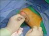
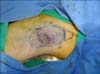
The patients were placed in supine position with their arms abducted to about 110°. Axillary hair was shaved before surgery, and the axillary hairline and 1 cm beyond the outline were marked on both sides. After routine sterilization of the area, an anesthetic containing 0.5% lidocaine solution (1 ml of 1:100,000 epinephrine, 10 ml of 2% lidocaine, and 30 ml normal saline) was injected on each side (20 ml). An incision of about one-third of the length of the widest transverse diameter was made on the axillary crease. The loosened layers between the dermis and subcutaneous tissue containing the apocrine glands were carefully undermined with iris scissors, and then the apocrine glands were separated from the skin. The remaining few apocrine glands attached to the dermis were removed by everting the thin flap, if necessary, while protecting the subdermal vascular plexus. Concurrently, any suspected hemorrhagic spots were immediately coagulated with an electrosurgical pencil (Valleylab/Covidien, Boulder, CO, USA). Then, the laser cannula was inserted into the dermal-subdermal junction through the incision line. The area of transcutaneous illumination indicated the positioning of the tips. The pulsed Nd:YAG laser (Accu-Sculpt; Lutronic, Goyang, Korea) was applied with a 1,444-nm wavelength, 40-Hz frequency, and 15-mJ energy (working at 6 W) (Fig. 1). In the authors' experience, these parameters produce the best results but must be adjusted according to situation. The apocrine glands within the marked area were destroyed by means of laser irradiation at intervals of 0.5 cm within 2 s. The total accumulated energy used in each axilla ranged from 1,800 to 2,100 J/cm2, depending on sweating intensity and the dimension of the treated area. The position and depth of the cannula tip were controlled through transcutaneous guidance with a red helium-neon light. The incision was sutured with 5~0 nylon (Fig. 2). The total procedure time was about 60 min. Immediately after treatment, cold normal saline bags were applied for about 5~10 min to minimize postoperative edema or discomfort, and to decrease the risk of possible skin damage or burning. Postoperatively, a small compressive dressing was applied for 2~3 days and the patients were advised to restrict their arm movements for 3 days. After the irradiation treatment, we followed all patients on postprocedure days 1, 7, 30, 60, 120, and 180. Relapse of osmidrosis was concluded when both the patient and the dermatologist were aware of the recurrence of malodor, or when no relevant improvement has been observed.
DISCUSSION
Many treatment modalities have been developed for axillary osmidrosis1. Those treatments are classified as either surgical or nonsurgical. A variety of surgical interventions have been used to treat axillary osmidrosis, including the removal of axillary tissue through minor skin excision and subcutaneous curettage, liposuction curettage, endoscopic sympathectomy, carbon dioxide laser vaporization, and ultrasound-assisted suction with skin incision. It is well known that surgical treatment, which removes subcutaneous tissue including the apocrine glands, provides the best results among the known procedures. However, it carries a high risk of adverse effects such as hemorrhage, hematoma, infection, scars or pain in the treated areas, and limitation of movement. The 1,444-nm lipolysis laser, used in facial lipoplasty and laser liposuction, has been recently introduced for use in apocrine gland removal. Thus far, subdermal coagulation treatment with a 1,444-nm Nd:YAG laser is a potentially less invasive and effective therapy for axillary osmidrosis2. However, this method may have a higher recurrence rate (~11%) than the surgical method2. This highlights the need for a combination of the surgical and nonsurgical methods. To maximize the advantages of surgery and minimize the risk of complications and adverse effects, we combined subcutaneous tissue removal and photothermal coagulation with a 1,444-nm Nd:YAG laser. Byeon et al.3 demonstrated that the apocrine glands in the axilla are mainly distributed in the center of the axillary region, especially within 6×3 cm. Therefore, we removed as many apocrine glands in the center of the axillary region as possible. Current nonsurgical methods involve the use of 1,064-nm pulsed Nd:YAG laser, as introduced by Kim et al.4 However, the 1,444-nm Nd:YAG laser has a greater lipolytic effect than the 1,064-nm Nd:YAG laser5. We therefore hypothesized that apocrine glands in fatty tissues would be more specifically destroyed by the 1,444-nm laser. From our results, the apocrine glands in the periphery of the axillary region were destroyed by irradiation with the 1,444-nm Nd:YAG laser.
All three patients exhibited no relapse of axillary osmidrosis and were satisfied with the treatment results. Two patients developed ecchymosis that resolved within several days. There were no severe complications including hematoma, granuloma, and postoperative infection. The combination of subcutaneous tissue removal and interstitial laser photothermal coagulation with a 1,444-nm Nd:YAG laser could be an effective treatment for cases of mild to moderate axillary osmidrosis, as the method is easy and has a minimal postoperative care requirement that allows patients to quickly return to their daily activities. Therefore, this method may be a less invasive and effective alternative therapy for axillary osmidrosis.
 XML Download