

 PDF
PDF ePub
ePub Citation
 Print
Print


INTRODUCTION
Vitiligo is an acquired skin disorder that is characterized by well-defined white patches caused by the disappearance of melanocytes in the epidermis1. There are many medical therapies, such as systemic and topical corticosteroid, photochemotherapy with psoralen plus ultraviolet-A, narrow-band ultraviolet-B, topical calcineurin inhibitors, and monochromatic eximer laser, are available for the treatment of this condition1. However, in cases of recalcitrant and stable vitiligo, surgical therapy has provided higher repigmentation rates than medical therapy1. Among the many surgical therapies, suction blister epidermal grafting (SBEG), which introduces active melanocytes to the lesion sites, is an established technique for the treatment of stable vitiligo2. Firm fixation of the graft on the recipient site is considered an important factor in SBEG treatment3.
Fibrin glue is a human plasma-derived material that has a hemostatic effect, helps in graft taking, and may have a protective effect against infection and provide an optimal environment for wound healing4. It is now approved by the Food and Drug Administration for multiple indications, including for hemostasis in a wide variety of surgical specialties and for colon sealing during colostomy closure5. Furthermore, it is widely used as an adhesive in plastic and reconstructive surgery, such as for skin graft attachment in burn wound grafting and in split-thickness skin grafts4. However, fibrin glue fixation for SBEG has not been studied in the treatment of stable vitiligo.
In this article, we report two cases of stable vitiligo treated with SBEG with fibrin glue fixation at sites where graft fixation is difficult. We suggest that fibrin glue fixation can be an alternative method for improving the result of SBEG.
CASE REPORT
Case 1
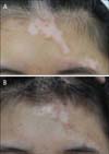
A 16-year-old female patient presented with well-defined 1~3 cm vitiliginous patches with poliosis on the forehead and frontal scalp area (Fig. 1A). One year ago, whitish patches with poliosis had developed on her forehead and frontal scalp area. She was treated with ultraviolet targeted phototherapy (Dualight; TheraLight Inc., Carlsbad, CA, USA) for 1 year; however, she showed no significant response to phototherapy. Therefore, we decided to try SBEG with fibrin glue fixation.
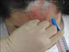
We created skin blisters by using a suction pump (HLSC9870; Hanlim, Seongnam, Korea) on the medial aspect of the thigh. After 2 hours of suction at 400 mmHg, small vesicles started to form, which enlarged to 1×1 cm in size; then, the blisters were carefully removed. The recipient sites were prepared by shaving the hairs on the forehead and frontal scalp area, and resurfacing with a CO2 laser. The graft was carefully implanted to the denuded recipient site, and approximately 1 ml of fibrin glue (Beriplast P; Aventis Behring, Marburg, Germany) was applied on the marginal area of the graft (Fig. 2). A porous silicone wound contact layer (Mepitel; Mölnlycke Healthcare, Goteborg, Sweden) was placed on the grafts, and simple compressive dressing with dry sterile gauzes was applied.
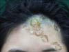
The dressings were removed after 7 days. At that time, firm fixation of the donor tissues was observed even in the hair-bearing areas (Fig. 3). Targeted phototherapy was restarted at 4 weeks after the SBEG, and about 50% repigmentation of the recipient areas was achieved after 6 months (Fig. 1B). No adverse effects such as scarring, pain, post-inflammatory hyperpigmentation, and Koebner phenomenon were observed in the patient.
Case 2
A 32-year-old woman presented with 1~2.5 cm hypopigmented patches on the chin (Fig. 4A). Depigmented patches developed on her chin about 10 years ago. Targeted phototherapy (Dualight) was given twice weekly for 4 years; however, no significant improvement was observed.
We performed SBEG in the same manner as in Case 1, and the grafts were fixed by using fibrin glue. After 7 days, a good graft uptake was observed and no adverse effects were noted. Treatment with a 308-nm xenon chloride eximer laser (XTRAC Laser; PhotoMedex Inc., Montgomeryville, PA, USA) was started at 4 weeks after the grafting, and complete repigmentation of the recipient areas was achieved after 18 weeks (Fig. 4B).
DISCUSSION
Although many medical therapeutic modalities have been improved recently, only limited successes in the treatment of vitiligo have been reported6. For patients who have recalcitrant and stable vitiligo, various surgical therapies can be applied, such as SBEG, split-thickness grafting, punch skin grafting, and transplantation of cultured melanocytes7. According to the literature, split-thickness grafting has a high success rate (>50% stable repigmentation for at least 6 months) of 78%~91%, which is comparable to that of SBEG. However, the procedure is difficult to perform in specific areas such as the eyelids, lips, and genitalia8,9. Various adverse effects, including hyperpigmentation, milia formation, curling of the graft, scar formation at the recipient site, and scar or keloid formation at the donor site, were also reported2. Punch skin grafting is an effective method for the treatment of stable vitiligo; however, it can also induce scarring and a cobblestone appearance7. Transplantation of cultured melanocytes can provide a new source of melanocytes for larger vitiligous lesions; however, this method is not cost-effective7.
SBEG is an established technique for the treatment of recalcitrant and stable vitiligo2. However, graft fixation is difficult in mobile areas such as the hands, foot, joints and cutaneous folds, and hair-bearing areas10. To overcome such difficulties, various methods have been attempted for the fixation of donor tissue7,11,12. The placement of stay suture is time consuming and carries the risk of tearing13. A film dressing can easily detach from cutaneous folds or hair-bearing areas, and the adhesive itself can cause graft detachment because it stick to the graft but not to the surrounding skin13. The splint can only be applied to the extremities; however, this is inconvenient for patients.
We report the nonconventional use of fibrin glue for fixing grafts. Fibrin glue is a topical hemostat, sealant, and adhesive that imitates the final stages of the coagulation cascade when a solution of human fibrinogen is activated by thrombin. Fibrin glue has a fibrinogen component and a thrombin component, both produced from human plasma4,5,14. Fibrin glue has been shown to improve the taking of skin graft, promote hemostasis, and confer a protective effect against bacterial infection by improving phagocyte motility in the fibrin and saturating the bacterial proteolytic enzymes by the action of the exogenous fibrin4,14. However, there is a potential risk of hypersensitivity and transmission of viral diseases (e.g., hepatitis A, B, and C; human immunodeficiency virus; Epstein-Barr virus; and cytomegalovirus)14. However, donor screening, heat treating, and use of a solvent/detergent suspension have made fibrin glues safe from viral transmission4. In a report on a large number of clinical applications, there were no adverse effects such as viral infection were observed after fibrin glue fixation14,15.
Fibrin glue seems to be an effective fixation method in SBEG. In our cases, simple dressing with nonadhesive porous silicone and dry gauzes were applied after SBEG with fibrin glue fixation. Both patients achieved >50% stable repigmentation for at least 6 months2. Our patients were satisfied with the treatment results, and no serious adverse effects were observed. Fibrin glue serves as a bioprotective film, resulting in reduced bacterial infection and conferring a protective effect by creating a physiological dressing that contributes to wound healing.
The use of fibrin glue in SBEG has some limitations. First, fibrin glue may interrupt graft attachment by permeating into the space between the graft and the underlying tissue. Second, we tried the treatment in an insufficient number of cases (i.e., two patients); therefore, despite the good results in these cases, further studies are needed. Finally, the high cost of the fibrin glue also needs to be taken into account.
Therefore, the application of an epidermal graft with fibrin glue can be an alternative treatment for stable vitiligo.

 XML Download