Abstract
Myiasis is a parasitic infestation of vertebrate animal tissues due to maggots of two-winged flies (Diptera) that feed on living or necrotic tissue. Dermatobia hominis occurs widely in tropical parts of Latin America; it is the most common cause of furuncular myiasis in this region. The continuous increase in international travel has increased the possibility of observing this pathology outside endemic countries, especially in travelers returning from the tropics. If clinicians are aware of the possibility of the disease and its treatment options, this dermatosis can be easily managed. However, diagnostic delay is very common because the disease is often misdiagnosed as a bacterial skin infection. Here, we report 2 cases of furuncular myiasis caused by D. hominis in travelers returning to Italy from Latin America. Surgical and noninvasive treatment approaches are also described.
Myiasis is a parasitic infestation of vertebrate animal tissues caused by maggots of two-winged flies (Diptera) that feed on living or necrotic tissue1.
We report two cases of furuncular myiasis caused by Dermatobia hominis in travelers returning to Italy from Latin America.
On October 5, 2009, a 39-year-old Brazilian woman currently living in Italy was referred to our outpatient clinic with 3 boil-like lesions: 2 were located on the scalp, and 1 on the right gluteus (Fig. 1). The woman had travelled through southeastern Brazil (Coronel Fabriciano, Minas Gerais) to visit her relatives for 1 month and returned on September 12. The nodules appeared at the beginning of September when she was in Brazil a few days after horse riding in a forest near a native village. The woman did not suffer any trauma, insect bites, or any other kind of injury at the lesion sites. After returning to Italy, she was treated with an antibiotic for 1 week by her Italian general practitioner, but the nodules did not stop growing.
Clinical examination confirmed the presence of 3 furuncular lesions approximately 1.5 cm in diameter with minimal perilesional erythema and a small central orifice where a whitish mobile body was visible. Her body temperature was 36.5℃, and right retroauricular tender lymphadenopathy was the only relevant finding on physical examination. Considering her history, the lesions were suspected to be furuncular myiasis.
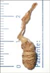
When we communicated our diagnostic impression to the patient, she began to be very frightened and anxious. First, we tried to asphyxiate the maggots by using petroleum jelly in order to force it to get out, but we failed. As the patient was becoming increasingly anxious, we decided to perform surgery to speed the larval extraction. We made an incision on the nodules after local anesthesia with lidocaine. Using pincers, we extracted a 3rd instar larva of D. hominis (Fig. 2) from each nodule. The procedure was complicated by the several concentric rows of backward-projecting spines around the maggots, which made the dislodgement quite difficult. The maggot in the nodule of the gluteus was accidentally crushed during the procedure. Therefore, very careful debridement was performed and open-packing medication was administered to avoid an allergic or foreign body reaction, or even a secondary infection. Amoxicillin/clavulanic acid (1 g q12 h orally for 5 days) was prescribed because of the presence of retroauricular tender lymphadenopathy and the procedural complication. The wounds normally healed in the following days.
On November 28, 2011, a 41-year-old Italian man who was fond of adventure travel to Latin America presented to our clinic with a single furuncular lesion in the skin of the interscapular region (Fig. 3). He returned from a 1-month trip to Bolivia on October 20. He precisely remembered that the skin lesion appeared as a small itchy papule after a bite from an unidentified insect while he was resting in a hammock on October 18 in Madidi National Park (upper Amazon river basin). In the following days, the papule evolved into a nodule with a small punctum through which serosanguineous brownish fluid discharged. The nodule started to grow slowly and did not heal despite daily dressing delivered by a nurse friend. The patient did not complain of any other symptoms except sporadic paroxysms of itching in the skin involved. When he came to visit our clinic, the nodule was approximately 1.5 cm and the small central punctum revealed a whitish body moving back and forth. Soft tissue ultrasonography showed a 1.7×1.2-cm hypoechoic lozenge-shaped formation in the context of the skin, with a moving upper part without any color Doppler signal (Fig. 4). We placed a 1-cm-thick layer of petroleum jelly on the nodule. After about 45 min, the larva inside the nodule began to emerge from the gel; at that moment, we squeezed the skin to eject the parasite, but the larva suddenly returned to hole. After another 15 min, the larva reemerged; this time, we quickly grabbed the emerging part with pincers and extracted an entire third instar larva of D. hominis (Fig. 5) by using moderate traction. The posterior of the maggot was partially damaged, where it was held to be extracted. After the extraction, we washed the remaining fistula with hydrogen peroxide. We prescribed only local medication and not post-procedural antibiotic treatment. The lesion healed in the following days.
Myiasis is a parasitic infestation of vertebrate animal tissues caused by maggots of two-winged flies (Diptera) that feed on living or necrotic tissue1,2,3. D. hominis is widespread in tropical parts of Latin America and is the most common causative species of furuncular myiasis in this region; other species are involved in other tropical and subtropical regions1. D. hominis larvae are obligate parasites that must develop in living tissues1. The main host is livestock, and humans are incidental hosts1. Female adult D. hominis lay their eggs on vector insects such as mosquitoes or flies2. The eggs hatch when the vector settles on a warm-blooded animal2. If transmitted by a biting insect such as a mosquito, the larva can enter the skin directly through the bite, which was probably the case in patient 22. Dermatobia eggs can also be transmitted by non-biting flies of the family Muscidae, which feed on liquid secretions such as sweat4. In such cases, the larvae can enter the skin by other means such as through hair follicles, which was probably the case in patient 12. The larva remains in the subdermal cavity for 5~10 weeks; when it matures, it will emerge from the skin, drop to the soil, and pupate1.
Myiasis represents 3.5%~9.3% of dermatosis cases in travelers returning from the tropics5. However, only few cases of furuncular myiasis due to D. hominis have been reported in Italy (Table 1)6,7,8,9,10,11,12,13,14. If clinicians are aware of the existence of the disease, the diagnosis can be suspected on the basis of a history of recent travel to the tropics and the presence of a furuncular lesion that does not heal with antibiotic treatment. However, the diagnosis is often delayed for more than 1 month from the last exposure15, probably because the maggot takes that long to grow such that the mobile spiracle is visible in the center of the nodule. Ultrasonography of the lesion can likely detect the movements of the maggot and confirm the diagnosis earlier. The first-line treatment is based on removal of the maggot by using noninvasive techniques5. Among noninvasive techniques, a very wide range of successful procedures have been reported; occlusion with petroleum jelly, pork fat, nail polish, tobacco tar, or bacon strips can cause the maggot to migrate to the surface, making extraction with forceps easier16. Moreover venom extractor to aspirate the larva and injection of lidocaine in to the lesion to increase the pressure and cause expulsion of the maggot have been used17,18. If noninvasive procedures fail, sterile surgical incision of the nodule and extraction of the maggot is indicated; care must be taken to avoid breaking the maggot to avoid foreign body or allergic reactions5. Besides larval extraction, all myiasis wounds must be cleaned and conservatively debrided, and tetanus immunization assessed and provided if needed5. Antibiotics are usually not recommended in the absence of secondary infection5. Our experience shows that the maggots can be easily damaged, so great attention should be paid when performing both invasive and noninvasive extraction techniques.
Figures and Tables
 | Fig. 2Case 1. Two of the three third instar maggots extracted; the third was crushed during the procedure. |
 | Fig. 3Case 2. Furuncular lesion in the skin of the interscapular region with visible maggot spiracle. |
 | Fig. 4Case 2. Soft tissue ultrasound showing a hypoechoic lozenge-shaped formation in the context of the skin. |
References
1. Robbins K, Khachemoune A. Cutaneous myiasis: a review of the common types of myiasis. Int J Dermatol. 2010; 49:1092–1098.

2. Maier H, Hönigsmann H. Furuncular myiasis caused by Dermatobia hominis, the human botfly. J Am Acad Dermatol. 2004; 50:2 Suppl. S26–S30.

4. Leite RC, Rodríguez Z, Faccini JL, Oliveira PR, Fernandes AA. First report of Haematobia irritans (L.) (Diptera: Muscidae) as vector of Dermatobia hominis (L.jr.) (Diptera: Cuterebridae) in Minas Gerais, Brazil. Mem Inst Oswaldo Cruz. 1998; 93:761–762.

6. Rizzo G, De Vito D, Rizzo C. A case of cutaneous myiasis caused by Dermatobia hominis. Parassitologia. 1998; 40:335–337.
7. Veraldi S, Gorani A, Süss L, Tadini G. Cutaneous myiasis caused by Dermatobia hominis. Pediatr Dermatol. 1998; 15:116–118.

8. Guidi B, Olivetti G, Sbordoni G, Garcovich A. Guess what! Diagnosis: cutaneous myiasis due to dermatobia hominis. Eur J Dermatol. 2001; 11:259–260.
9. Matera G, Liberto MC, Larussa F, Barreca GS, Focà A. Human myiasis: an unusual imported infestation in Calabria, Italy. J Travel Med. 2001; 8:103–104.
10. Romano C, Albanese G, Gianni C. Emerging imported parasitoses in Italy. Eur J Dermatol. 2004; 14:58–60.
11. Bongiorno MR, Pistone G, Aricò M. Myiasis with Dermatobia hominis in a Sicilian traveller returning from Peru. Travel Med Infect Dis. 2007; 5:196–198.

12. Varani S, Tassinari D, Elleri D, Forti S, Bernardi F, Lima M, et al. A case of furuncular myiasis associated with systemic inflammation. Parasitol Int. 2007; 56:330–333.

13. Calderaro A, Peruzzi S, Gorrini C, Piccolo G, Rossi S, Grignaffini E, et al. Myiasis of the scalp due to Dermatobia hominis in a traveler returning from Brazil. Diagn Microbiol Infect Dis. 2008; 60:417–418.

14. Veraldi S, Francia C, Persico MC, La Vela V. Cutaneous myiasis caused by Dermatobia hominis acquired in Jamaica. West Indian Med J. 2009; 58:614–616.
15. Schwartz E, Gur H. Dermatobia hominis myiasis: an emerging disease among travelers to the Amazon basin of Bolivia. J Travel Med. 2002; 9:97–99.

16. Diaz JH. The epidemiology, diagnosis, management, and prevention of ectoparasitic diseases in travelers. J Travel Med. 2006; 13:100–111.


 PDF
PDF ePub
ePub Citation
 Print
Print


 XML Download