

 PDF
PDF ePub
ePub Citation
 Print
Print


INTRODUCTION
Skin aging includes pigmentary alterations, wrinkling, thinning, and loss of elasticity owing to both genetic and environmental factors. Various medical treatments and topical cosmeceuticals are used to treat symptoms of aging. However, the results have been unsatisfactory thus far.
Endothelial precursor cells (EPCs) differentiated from human embryonic stem cells (hESCs) demonstrated improvement of blood perfusion in damaged tissues secreting high levels of growth factors and cytokines1,2. Conditioned medium (CM) of hESC-derived EPCs (hESC-EPCs), which comprises several growth factors and cytokines, significantly improved the proliferation and migration of dermal fibroblasts and epidermal keratinocytes as well as increased collagen synthesis in fibroblasts2. In this respect, growth factors may be beneficial for reducing signs of skin aging3. Some growth factors also exhibit a whitening effect by inhibiting melanogenesis4. However, the beneficial role of growth factors for skin rejuvenation has only recently been to be studied5,6,7, and no controlled clinical trials have been performed.
Hydrophilic molecules larger than 500 Da have poor penetration through the stratum corneum8,9. Most growth factors are large hydrophilic molecules greater than 20 kDa; therefore, they are unlikely to penetrate the epidermis in measurable quantities to produce pharmacologic effects. This 12-week double-blinded randomized split-face study was performed to investigate the effects of the secretory factors of hESC-EPC on aged skin in Asians. Microneedling was used to enhance the skin penetration of hESC-EPC CM. An in vitro experiment to confirm the cutaneous absorption of hESC-EPC CM after microneedling was also performed. In particular, this study used diverse noninvasive skin-measuring devices to objectively assess changes in the biophysical properties of the skin following hESC-EPC CM treatment.
MATERIALS AND METHODS
Preparation of conditioned medium and multiplex cytokine assay
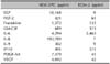
We used commercially available CM of hESC-EPCs, which were generated as described previously10. The multiplex cytokine array was performed using the Milliplex and Luminex systems (Millipore; Luminex Corp., Austin, TX, USA) with concentrated CM; this system can be used to analyze all or any combination of cytokines and chemokines in tissue/cell lysate and culture supernatant samples by using a microbead-based tagging system2. Endothelial growth medium-2 (EGM-2; Lonza, Walkersville, ML, USA) was used as control medium. The multiplex cytokine analysis of CM revealed that hESC-EPCs strongly expressed several growth factors including epidermal growth factor (EGF), fibroblast growth factor-2 (FGF-2), fractalkine, granulocyte macrophage colonystimulating factor (GM-CSF), interleukin-6 (IL-6), plateletderived growth factor-AA (PDGF-AA), and vascular endothelial growth factor (VEGF) (Table 1).
Cutaneous absorption experiment
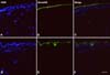
To confirm transdermal absorption, proteins in hESC-EPC CM were labeled by using the Alexa Fluor 488 Protein Labeling Kit (Invitrogen Co., Carlsbad, CA, USA) according to the manufacturer's protocol. Briefly, Alexa Fluor 488 reactive dye was mixed with 1 mg protein in 1.5 ml of 0.1 M sodium bicarbonate buffer. After 1 hour, the unreacted dye was separated by using a purification resin column. Female miniature pig skins (Medi Kinetics Micro-pigs; Medi Kinetics Co., Ltd, Busan, Korea) were microneedled using 0.25-mm-long microneedles. Fluorescence dye-protein conjugates were subsequently applied to the skin and incubated for 1 hour. Tissues were immediately embedded in frozen sectioning compound (Leica Microsystems GmbH, Wetzlar, Germany) in liquid nitrogen. The sections were approximately 16 µ thick and were dried overnight at room temperature. The slides were washed several times with PBS and incubated with Fluoroshield Mounting Medium with DAPI (ImmunoBio-Science Co., Mukilteo, WA, USA). Images were captured by a Nikon fluorescence microscope (Nikon, Tokyo, Japan) at excitation wavelengths of 405 nm and 488 nm and merged.
Study design and participants
Twenty-five participants were recruited for this prospective randomized controlled observer-blinded split-face study. Participants were 41~64 years old (mean, 51.6 years) and had Fitzpatrick Skin Type III or IV. The exclusion criteria were as follows: use of bleaching creams, history of any skin rejuvenation treatment within 6 months, history of keloids, and active eczema. The study protocol and informed consent form were submitted to and approved by the CHA University institutional review board (PBC10-062).The participants were informed of the benefits, risks, and possible complications of the treatment before enrollment; all provided informed consent prior to participation.
The left and right sides of the face of each participant were randomly assigned to treatment with microneedling alone (control) or microneedling plus hESC-EPC CM. The randomization procedure involved sealed envelopes numbered 1~25 in which the allocation was indicated. The randomization was based on a computer generated random list (GraphPad Software Inc., La Jolla, CA, USA) created by an independent cooperator, and envelopes were opened in ascending order. All participants and 2 dermatologists assessing outcomes were blinded until all the participants finished final assessments.
Participants received 5 treatments at 2-week intervals. First, the face was anesthetized by topical 4% lidocaine cream (LMX4; Ferndale Laboratories Inc., Ferndale, MI, USA) approximately 30 minutes before the procedure. The face was cleansed with a mild soap and 70% alcohol. For the microneedling alone treatment, 1.5 ml of normal saline was painted on the skin, and 2 passes of microneedling with dermarollers (0.25-mm DTS roller; TCellBio, Seoul, Korea) were performed. The endpoint of treatment was the presence of uniform erythema over the face. For the microneedling plus hESC-EPC CM treatment, 1.5 ml of hESC-EPC CM was painted on the face and microneedling was performed in the same manner. An epidermal cooling device (CARESYS; Danil SMC, Seoul, Korea) was used to relieve pain and erythema after microneedling therapy.
Clinical assessments
Participants were assessed at baseline and 2 weeks after final treatment (12 weeks). Photographs taken by a digital camera (Nikon D90; Nikon) were obtained at each visit. For self-assessment, participants answered questionnaires regarding efficacy and adverse events 12 weeks after study initiation. The questionnaires included grading of overall treatment satisfaction from 0 (dissatisfied) to 5 (most satisfied). In addition, participants were asked to report any side effects during the study. Objective clinical assessments consisted of 2 dermatologists blinded to the study design and treatment comparing pre- and posttreatment photographs separately on each side of the face. The evaluations were graded by quartile as follows: grade 1, 0%~25%, minimal to no improvement; grade 2, 26%~ 50%, moderate improvement; grade 3, 51%~75%, marked improvement; and grade 4, 75%~100%, near total improvement.
Non-invasive objective skin color measurements
Prior to all measurements, participants were acclimatized to a temperature- (20℃) and humidity-controlled (40%) room, and the instruments were calibrated according to the manufacturer's instructions. A narrow-band simple reflectance meter, Mexameter (MX18; Courage+Khazaka Electronic GmbH, Köln, Germany), was used to quantitatively evaluate color changes after treatments; this instrument uses arrays of light emitting diodes (LEDs) that emit light at 3 defined wavelengths: 568 (green), 660 (red), and 880 nm (infrared). The melanin index (MI) and erythema index (EI) were measured in triplicate on the same malar area on each side of the face, and mean values were used for analysis.
Non-invasive objective wrinkle measurements
To evaluate the effects of treatments on collagen regeneration, each participant's periorbital wrinkles were objectively measured by using a skin replica and microrelief instrument (Visiometer SV600; Courage+Khazaka Electronic GmbH) at baseline and 12 weeks. The Visiometer SV600 can measure skin roughness and the depth of furrows by measuring the light transmission through a very thin skin replica. The roughness parameters investigated in this study were R2 (maximum roughness) and R3 (average roughness).
RESULTS
Cutaneous absorption experiment
The proteins in hESC-EPC CM labeled with the Alexa Fluor 488 Protein Labeling Kit were visualized in both the epidermis and dermis (Fig. 1).
Clinical assessments
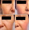
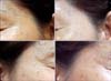
All 25 participants completed the 12-week study protocol. No serious adverse events were encountered. Mild pain and temporary erythema during and after treatments were tolerable in all participants. The only minimal adverse event reported in one participant was mild desquamation, which resolved spontaneously within 1 week. Participants' overall satisfaction scores for microneedling alone and microneedling plus hESC-EPC CM were 2.72±1.45 and 3.25±1.26, respectively (p<0.05; Table 2). The mean grades of objective clinical improvement of pigmentation based on photographs for microneedling alone and microneedling plus hESC-EPC CM were 1.32±0.62 and 1.54±0.57, respectively (p<0.05; Table 2). The mean grade of objective clinical improvement of wrinkles based on photographs for microneedling alone and microneedling plus hESC-EPC CM were 1.49±0.48 and 1.92±0.42, respectively (p<0.05; Table 2). Representative photographs showed greater improvements in wrinkles and dilated pores following microneedling with hESC-EPC CM than microneedling alone (Fig. 2, 3).
Pigmentation improvement
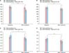
At baseline, the MI determined by Mexameter revealed no statistically significant difference in pigmentation between microneedling alone and microneedling plus hESC-EPC CM (p=0.15). However, treatment with microneedling plus hESC-EPC CM resulted in a significantly greater decrease in the MI than microneedling alone (p<0.05, Fig. 4A). The mean MI of the sides treated with microneedling alone decreased from 143±11.1 at baseline to 136±12.8 two weeks after the final session (p=0.052). Meanwhile, the mean MI of the microneedling plus hESC-EPC CM sides decreased significantly from 138±14.2 at baseline to 113±12.1 two weeks after the final session (p<0.05), demonstrating the efficacy of hESC-EPC CM for skin lightening.
Erythema improvement
Treatment with microneedling plus hESC-EPC CM significantly decreased the EI from 271±24.2 at baseline to 243±21.1 two weeks after the final session (P<0.05, Fig. 4B). The mean EI of the sides treated with microneedling alone decreased from 268±25.1 at baseline to 255±29.8 two weeks after the final session (p>0.05).
Wrinkle improvement
Treatment with microneedling plus hESC-EPC CM resulted in a significantly greater decrease in the R2 and R3 values measured by Visiometer than microneedling alone (p<0.05; Fig. 4C, D). The mean R2 value of the sides treated with microneedling alone decreased from 0.52±0.07 at baseline to 0.50±0.1 two weeks after the final session, but the changes were not significant (p>0.05). Of note, the R2 values of the sides treated with microneedling plus hESC-EPC CM decreased significantly from 0.58±0.1 at baseline to 0.46±0.09 two weeks after the final session (p<0.05). Similarly, the mean R3 value of the sides treated with microneedling alone decreased from 0.38±0.06 at baseline to 0.34±0.1 two weeks after the final session (p=0.51). Meanwhile, the mean R3 values of the sides treated with microneedling plus hESC-EPC CM decreased significantly from 0.4±0.1 at baseline to 0.31±0.06 two weeks after the final session (p<0.05).
DISCUSSION
Skin aging is mediated by the effects of both the natural aging process (i.e. intrinsic aging) and environmental factors (i.e. extrinsic aging) on cellular and extracellular components. Cell-based therapies using the body's own stem cells and growth factors have recently been used as an alternative therapeutic strategy to repair damaged tissue, including skin rejuvenation. Stem cells may exert their beneficial effects on tissue regeneration through complex paracrine mechanisms in addition to their proposed direct cellular effect11. Furthermore, stem cells synthesize and secrete a variety of extracellular matrix proteins, cytokines, growth factors, and other bioactive proteins that contribute to the healing process; the local environment created by these secreted factors may govern the fate and function of individual stem cells11,12,13.
A previous study revealed that hESC-EPC CM accelerates wound healing and increases the tensile strength of wounds after topical treatment and subcutaneous injection1. In vitro, hESC-EPC CM significantly improved the proliferation and migration of dermal fibroblasts and epidermal keratinocytes, and also increased collagen synthesis by fibroblasts2. Analysis of hESC-EPC CM by using a multiplex cytokine array system revealed that hESC-EPCs secrete cytokines and chemokines such as EGF, bFGF, fractalkine, GM-CSF, IL-6, IL-8, PDGF-AA, and VEGF, which are important in normal angiogenesis and wound healing2. In addition, conditioned media from adipose-derived stem cells (ADSC CM) have shown to inhibit melanogenesis by downregulating tyrosinase and tyrosinase-related protein-1 expression in B16 melanoma cells, demonstrating the whitening effects of ADSCs14. Similarly, hESC-EPC CM inhibited melanogenesis in B16 melanoma cells (unpublished data). Therefore, we hypothesized that hESC-EPC CM improves the signs of skin aging such as wrinkles and pigmentation. The present study demonstrated that 5 sessions of hESC-EPC CM application significantly improved skin pigmentation and wrinkles.
Transdermal penetration and epidermal-dermal communication are important regarding the cutaneous application of growth factors for skin rejuvenation. Despite the 500 Da rule for the skin penetration of chemical compounds, there are several potential routes by which small quantities of large molecules can penetrate the stratum corneum, such as the follicular route15,16. Vaccines larger than 100 kDa were recently found to be able to exert an immunologic response when applied topically, probably because of the penetration of a very small amount of protein through intact skin17. Penetration into the uppermost layer of viable epidermal keratinocytes may produce a signaling cascade of growth factors that affects cells deeper in the dermis such as fibroblasts.
In the present study, microneedling was performed to enhance the skin penetration of hESC-EPC CM. Microneedling, which is a collagen-induction therapy, is a method that creates pinhole wounds using many microneedles. It stimulates wound healing and improves scars and wrinkles18. Moreover, microneedles have been used in transdermal and dermal drug delivery for more than a decade19. We confirmed that proteins in hESC-EPC CM can directly penetrate the epidermis when combined with 0.25-mm microneedling. Therefore, the presence of hESC-EPC CM in the dermis is expected to exert direct effects on the dermal extracellular matrix.
The main limitations of this study are the small number of participants and lack of the long-term follow-up after final treatment. However, the randomized split-face study design enhances the reliability of the data as it enabled us to obtain significant results with a relatively small group of participants.
Although there are reports of the roles of soluble factors of stem cells in photoaging, this is the first randomized controlled study, which demonstrates the efficacy of the soluble factors of stem cells on skin rejuvenation in vivo.

 XML Download