

 PDF
PDF ePub
ePub Citation
 Print
Print


INTRODUCTION
Skin infections with Pseudomonas aeruginosa and Escherichia coli occur more frequently in patients who are immunologically compromised, elderly, or who are in intensive care units (ICU). These patients may have other comorbidities, including deteriorated renal and hepatic function, administration of multiple other drugs, and complications from chronic diseases. Thus, the administration of systemic antibiotics to these patients has a higher risk of side effects. In addition, there is a narrow range of antibiotics that can be used to treat infection, because many strains of P. aeruginosa and E. coli show resistance to certain antibiotics.
Useful topical treatment for Gram-negative bacterial infection is limited. The use of topical antibiotics and antiseptics are also limited due to bacterial resistance and the inherent nature of treatment agents. For instance, some antibiotics/antiseptics contain superoxide-generating hydrogen peroxide. Further, potassium permanganate and chlorhexidine are cytotoxic and can inhibit the regeneration of cells for wound repair1,2.
Epigallocatechin gallate (EGCG) is a major catechin found in green tea extracts (GTE), and has remarkable antibacterial activity as well as anti-oxidant, anti-inflammatory, and anti-cancer effects. Further, EGCG can induce epithelial proliferation and differentiation3,4,5. EGCG is known to be active against Gram-positive bacteria4,6. It is also known that the minimum inhibitory concentration (MIC) of EGCG in Gram-negative bacteria appears to be 8- to 16-fold higher than that in Gram-positive bacteria6. The relatively lower antimicrobial activity of catechins against Gram-negative bacteria is due to the protection of the Gram-negative bacteria by the outer membrane and lipopolysaccharides.
This study was conducted to assess the possible use of EGCG and GTE as antibacterial agents in Gram-negative P. aeruginosa and E. coli, regardless of antibiotic resistance status.
MATERIALS AND METHODS
Bacterial strains
The studied bacterial strains were isolated from the skin wounds of patients admitted to the ICU at our University Hospital. Twenty-two strains in total were studied, and consisted of 10 P. aeruginosa strains, 10 E. coli strains, and 2 reference strains (P. aeruginosa ATCC 27853, E. coli ATCC 25922) for quality control. Cultured strains were kept at a turbidity matching a 0.5 McFarland standard and placed in each well with a final concentration of 2.5×10⁵ colony-forming unit (CFU) /ml.
Antibiotic susceptibility tests of the bacterial strains
Specimens from the skin wounds of the ICU patients were inoculated and cultured on blood agar medium and MacConkey agar medium for 24 to 48 hours at 35℃. The identification of bacterial isolates was performed manually and processed simultaneously using the ATB ID system (BioMériuex, Marcy l'Etoile, France). Antibiotic susceptibility was assessed using the disk diffusion method following the guidelines published by the Clinical and Laboratory Standards Institute (CLSI)7 for ampicillin, amikacin, aztreonam, cefoxitin, cefepime, cephalothin, cefotaxime, ciprofloxacin, ceftazidime, gentamicin, imipenem, piperacillin, piperacillin/tazobactam, and sulperazone. The results of the antibiotic susceptibility tests of isolated strains were compared with the results of EGCG and GTE susceptibility tests.
EGCG and GTE preparation
EGCG (Sigma, St. Louis, MO, USA) in powder form with 95% purity was dissolved in normal saline and serially diluted with cation-adjusted Mueller-Hinton broth (MHB) starting from a maximum concentration of 800 µg/ml to 0.4 µg/ml with 12 levels of dilution. Two grams of dried green tea leaves (product name: Halla; Amorepacific Corporation, Seoul, Korea) were brewed with 100 ml of boiling water for 10 min. After removing the tea leaves, the infusion was left for another 10 min and then cooled down to below 40℃. The green tea infusion was filtered twice using Whatman filter paper (Whatman International Ltd., Maidstone, UK). Final filtration by a filter with a pore size of 0.22 µm was performed to remove possible microbial contaminants. Crude GTE was diluted from undiluted extract in 12 dilution stages (1:1~1:2,048) by successively adding two-fold MHB.
Measuring minimal inhibitory concentration
Gentamicin (Sigma) and ciprofloxacin (Fluka, Saint Gallen, Switzerland) were used as reference antibiotics to assess the antimicrobial activity of EGCG and GTE against P. aeruginosa8. Ampicillin (Sigma) and ciprofloxacin were tested against E. coli. Ciprofloxacin was dissolved in 0.1 N HCl, and other antibiotics and EGCG were dissolved in normal saline. The antibiotics were serially diluted with cation-adjusted Muller-Hinton broth from a concentration of 256 µg/ml to 0.125 µg/ml, and 200 µl of the serially diluted antibiotics, EGCG or GTE was placed in a 96-well plate (Corning, Acton, MA, USA). E. coli and P. aeruginosa were inoculated in every well at a concentration of 2.5×10⁵ CFU/ml. The MIC was visually determined after 16~20 hours of incubation at 37℃ according to the broth microdilution methods suggested by the CLSI8.
RESULTS
Antibiotic susceptibility of the studied strains of P. aeruginosa and E. coli
1) P. aeruginosa
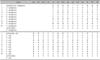
Five of the ten collected strains and the reference strain were susceptible to all of the tested antibiotics, while the other five strains showed multidrug resistance. These multidrug-resistant strains were commonly resistant to ciprofloxacin, gentamicin, piperacillin, and piperacillin/tazobactam. All ten of the studied strains were susceptible to imipenem and ceftazidime (Table 1).
2) E. coli
Only one of the collected E. coli strains and the reference strain were susceptible to all of the tested antibiotics. Nine of the ten strains were resistant to at least one antibiotic (up to nine antibiotics), although they did not show any similarities in their patterns of resistance. All of the E. coli strains were susceptible to cefoxitin, amikacin, imipenem, piperacillin/tazobactam, and cefoperazone/sulbactam (Table 1).
MICs of ciprofloxacin, gentamicin, EGCG, and GTE against P. aeruginosa
1) Three P. aeruginosa strains were resistant to ciprofloxacin. However, the growth of one strain was not suppressed, even at a concentration of 256 µg/ml. The reference strain and six of the studied P. aeruginosa strains were susceptible to gentamicin, with MICs around 1 µg/ml. The other four P. aeruginosa strains showed a high degree of resistance to gentamicin, even up to 256 µg/ml (Table 2).
2) The MIC of EGCG against six of the P. aeruginosa strains was 400 µg/ml, while the MIC of the other four strains and reference strain was 200 µg/ml. Two of the ten P. aeruginosa strains were susceptible an 8-fold dilution of crude GTE, whereas the other eight strains and the reference strain were susceptible at a 16-fold dilution (Table 2).
3) The MIC of EGCG and the dilution levels of GTE were not related to the degree of resistance of P. aeruginosa to ciprofloxacin and gentamicin.
MICs of ciprofloxacin, ampicillin, EGCG, and GTE against E. coli
1) Three of the ten strains of E. coli and the reference strain were susceptible to ciprofloxacin. The E. coli studied had a higher MIC and a higher incidence of resistance to antibiotics than P. aeruginosa. Eight of the E. coli strains were resistant to ampicillin, with uninhibited growth at a concentration of 256 µg/ml (Table 3).
2) The MIC of EGCG against all E. coli strains, including reference strain, was 400 µg/ml. In the case of GTE, the growth of the reference strain was inhibited at a 1:16 dilution. The MIC of GTE for four E. coli strains was an 8-fold dilution, whereas the MIC of the other six E. coli strains was a 4-fold dilution (Table 3).
3) The degree of resistance of the E. coli strains to ciprofloxacin and ampicillin was not related to the MIC of EGCG and GTE.
DISCUSSION
Skin and soft tissue infections with P. aeruginosa and E. coli have been an issue since both incidence and drug resistance are increasing. In healthy people, P. aeruginosa rarely causes infection; however, it poses a serious health risk in hospitals, where it is responsible for about 10% of in-hospital infections in immunocompromised patients, including those with cancer, diabetes, and hematological disorders, as well as patients undergoing transplant, receiving implants, and being treated with corticosteroids and antibiotics9. In Taiwan, it was reported that E. coli was responsible for almost one-fifth of skin and soft tissue infections10. In Europe, E. coli accounted for 10.8% of infections, and the corresponding rate was 7.2% for North America11. With the observed increase in the rate of bacterial resistance to an extended spectrum of antibiotics, clinical cures and the selection of active antibiotics for empirical treatment may be more difficult to achieve12.
A major component of green tea is a flavonol known as catechin13. (-)-Epicatechin (EC), (-)-epigallocatechin (EGC), (-)-epicatechin gallate (ECG), and EGCG are the four main catechins. In particular, EGCG is found only in green tea, comprising 40%~50% of green tea catechins and is thought to be primarily responsible for the antibacterial and bactericidal properties of green tea4,5,6,14,15. Gallocatechin and gallate are necessary moieties for antibacterial activity, with gallate-conjugated (-)-ECG and (-)-EGCG demonstrating more powerful antibacterial activity than non-gallate-conjugated (-)-EGC and (-)-EC14.
It has been reported that the bactericidal effect of EGCG is stronger in Gram-positive bacteria than in Gram-negative bacteria, owing to the different amounts of EGCG absorbed by the bacterial cells6,15,16. While the MIC of EGCG against Staphylococcus aureus, Staphylococcus epidermidis, Staphylococcus hominis, and Staphylococcus haemolyticus has been reported to be 50~100 µg/ml, the MIC of EGCG against Klebsiella pneumoniae, Salmonella typhi, and Proteus mirabilis is much higher (800 µg/ml)6. GTE has also shown various degrees of antibacterial activity, and a wide range of susceptibility against different strains of the same species6,17,18. One proposed mechanism for the bactericidal action of catechin is that the negatively charged EGCG combines with the positively-charged bacterial lipid polysaccharide membrane generating hydrogen peroxide (H2O2), which damages the bacterial membrane3,15. Gram-negative bacteria are generally more resistant to catechins than Gram-positive bacteria, due to the presence of strong negative charge of lipopolysaccharides on the exterior outer member of Gram-negative bacteria16. EGCG is known to have unique dual actions, and it protects human keratinocytes and fibroblasts against H2O2 by reversing the H2O2-induced decrease of superoxide dismutase (SOD) and glutathione peroxidase19,20.
The amount of EGCG or catechins in green tea differs depending on the product and extraction method. However, when green tea is infused in hot water for 3 min in a proportion of 1 g of leaves to 100 ml of water, the tea usually contains 250~280 mg of solids, of which 30%~42% are catechins3. When green tea or EGCG capsules are orally administered, only 0.2%~2.0% of the ingested EGCG is intestinally absorbed and appears in the blood17,18. Considering its low plasma concentrations and the reported MIC of EGCG against Gram-negative and Gram-positive bacteria, topical application of EGCG on infected lesions is more desirable than systemic administration, since the concentrations required to treat bacterial infections of the skin cannot be reached through drinking green tea.
Based on the previously reported proportion of catechins in green tea infusions, the estimated concentration of EGCG in the crude infusion of GTE prepared in this study was roughly 400~800 µg/ml21,22. At a GTE dilution level of 1:16, the EGCG concentration was about 25~50 µg/ml. Despite the low concentration of EGCG in the tested GTE, GTE showed antibacterial activity equivalent to 400 µg/ml EGCG. This can be explained through the synergistic antibacterial action of other polyphenols in GTE, such as ECG. Considering that even a 16-fold dilution of crude GTE showed effective antibacterial activity, GTE may be practical and economically feasible for use as an alternative for topical antibiotics or as dressing agents in clinical practices. Furthermore, the clinical application of catechin is plausible, since catechin is very stable under physical manipulations, such as freezing and heating, and can be refrigerated in an aqueous solution for over a month with good stability23,24.
The bacterial strains in this study were isolated from the ulcers and sores of long-stay patients admitted to the ICU. Thus, the incidence of multidrug resistant strains was higher than community acquired strains. However, bacterial strains showing multidrug resistance did not have the same pattern of resistance against EGCG or GTE, and showed susceptibility to EGCG or GTE independent of the antibiotic resistance status. Though the antibacterial effects of EGCG and GTE varied with the individual strains of bacteria, consistent levels of effectiveness were seen regardless of the susceptibility of bacteria to the reference antibiotics. Since EGCG exerts antibacterial effects through diverse mechanisms in vivo, the effective MICs of EGCG against P. aeruginosa and E. coli on skin lesions will be lower than the MICs seen in this study.
Better antimicrobial effects can be expected from GTE than EGCG, since GTE contains various types of catechins with antibacterial activity other than the four major catechins: EGCG, ECG, EGC, and EC. Furthermore, GTE is easily available and would be more cost-effective. Although more studies on their mechanisms of action are needed, EGCG and GTE have great potential for use as topical antimicrobial agents with systemic antibiotics to manage skin infections. Several experiments that tested the ability of EGCG to synergistically inhibit methicillin-resistant S. aureus with concomitant use of oxytetracycline, carbapenem, and ampicillin/sulbactam in vitro demonstrated that EGCG synergistically inhibited bacterial growth25,26,27. An in vivo study of chronic E. coli bacterial prostatitis rat model showed synergistic effects between an oral gavage of 300 mg/kg body weight of catechin concentrate and ciprofloxacin28.
The clinical application of EGCG and GTE is worth considering as a therapeutic in pursuit of overcoming the increasing antibiotic resistance of bacteria and further studies are needed29.


 XML Download