

 PDF
PDF ePub
ePub Citation
 Print
Print


INTRODUCTION
Skin cancer is the most common cancer in countries with predominantly fair-skinned populations. Basal cell carcinoma (BCC) is the most common type of skin cancer, usually occurring in the sun-exposed areas1,2. Its annual incidence is estimated at 2.75 million new cases worldwide3, with a projected annual increase of 10%, which may be due to the sun exposure and increased outdoor activities4,5. Despite the high prevalence of these tumors, there is a lack of reliable epidemiological data in some countries, including Iran. Although BCC is not an aggressive carcinoma and usually does not metastasize, its morbidity and the public health burden are significant, especially because it usually occurs on the head and neck area with a high recurrence rate3. BCCs are categorized according to the histological appearance. Major subgroup is nodular, but regarding aggressiveness and recurrence, infiltrating, micronodular, multifocal, superficial, morphoeiform and mixed histopathological patterns are significant as well3,6. Of the different treatment methods, surgery is the most common for the effectiveness7.
Recent studies show that BCC occurring in different anatomical sites may define specific clinical behavior and different etiologies8,9,10. According to literature, BCC subtypes occurring on different body sites are different from each other. For example BCC occurring on trunk is usually a superficial spreading form11.
Therefore, understanding the disease process, patterns of behavior and treatment options are necessary for plastic and dermatological surgeons. Indeed, assessment of relationship between tumor anatomical distribution and histological subtype may provide necessary knowledge for understanding pathogenesis of the disease, especially impact of sun exposure being the most important factor in the development. Estimates from the American Cancer Society suggest that there are more than two million cases of nonmelanoma skin cancer (NMSC) in the United States per year. The incidence of NMSC, which has increased over the past 20 years worldwide, may be related to higher levels of outdoor activities and sun exposure, changes in clothing style and improved skin cancer detection methods12,13. Raised healthcare concerns for tumors may be another reason for the increased incidence. Analysis of other epidemiological factors, such as geographic variation, age distribution and ethnicity, has also reinforced the theory that chronic sun exposure acts as a primary causative factor in NMSC. It would be of interest to assess whether a particular site has a preferential histologic subtype.
As there are no relevant data available concerning BCC in Iran, this study is designed to assess the relation between BCC subtypes and anatomical distribution in the Iranian patients.
MATERIALS AND METHODS
The study is approved by deputy of research, Tehran University of Medical Science. Among 3,400 patients referred to the Razi Dermatology Center (Tehran, Iran) from March 2007 to March 2010, 876 patients who were confirmed for BCC were enrolled in this study. Patients with recurrent tumors were also included, without the recurrences counting as separate cases. Samples were included on the basis of histopathological diagnosis of BCC, which was confirmed by two dermatopathologists.
Classification of tumor subtypes were done using the World Health Organization classification. Informed consent was obtained from all patients. Skin color types of all patients were Type III and IV of Fitzpatrick Classification Scale. Patients with medical conditions such as diabetes mellitus and those taking medication that compromised the immune system were excluded from the study. Demographic data with tumor location and histologic diagnosis were also obtained from all patients, along with the medical records.
Statistical analysis
The results are expressed as mean±standard deviation. Statistical analysis was performed using SPSS version 16.0.1 (SPSS Inc., Chicago, IL, USA). Statistical difference between proportions were determined by χ2 analysis. Numerical data were evaluated using analysis of variance, followed by Tukey's post hoc test. p<0.05 was considered as significant.
RESULTS
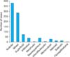
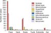
Among 876 patients, 544 (62%) were males and 332 (38%) females, with mean age of 63.0±12.1 years and 60.9±13.0 years, respectively. Of these patients, 457 underwent excisional biopsy, and 419 underwent punch biopsy. The largest diameter of lesions having undergone excisional biopsy had a mean of 2.42±1.85 mm. Among 876 patients, 379 (43.0%) presented with nodular subtype, 284 (32.4%) with mixed subtype, 27 with superficial and the 186 remaining presented with other subtypes (Fig. 1). For the lesions, 514 (58.6%) were on the face, 258 (29.5%) on scalp, 55 (6.3%) on ears, 21 (2.4%) on neck, 16 (1.8%) on trunk and 12 (1.4%) on the extremities, showing that 96.8% of the lesions were on the head and neck of the patients (Table 1). Among the 284 mixed BCCs, 79 were nodular and infiltrative, 59 were nodular and superficial, 49 were nodular and micronodular, 29 were nodular and adenoid and others were a combination of various BCC subtypes. Fifty-two percent of the mixed type BCCs were on the face, 37% on scalp, 8% on ear, 2% on neck and the remaining on trunk.
Histologic subtypes of BCC within the different anatomical sites are outlined in Fig. 2. There was no significant difference between male and female in BCC subtypes, but anatomical distribution of the tumors was different, in which scalp and ear BCC occurred less common in female than male. More than 65.0% of BCCs in female patients occurred on the face and 21.7% on the scalp, while the percentage in male patients were 52.2% and 33.8%, respectively (p=0.002). According to our results, different BCC subtypes occurred on different anatomical sites, with a statistically significant difference (p<0.001). The results showed that most BCCs on the trunk were superficial while most facial BCCs were nodular subtype (p<0.001). According to our results, most of the BCC subtypes occurred in patients between 40 to 80 years old and mostly on the face and scalp (p=0.04). Superficial subtype mostly occurred in younger patients than other subtypes (p=0.001). As mentioned above, the mean age of BCC occurrence in male patients was higher than in female patients (p=0.001). Infiltrative subtype presented with ulceration more frequently than other subtypes (p=0.002).
DISCUSSION
BCC is the most frequent cancer in humans14. Similar to other reports, present study showed that BCC occurred predominantly on the head and neck. Anatomical distribution of the subtype-specific rates showed that most of the trunk-arising BCCs were superficial, while most of the facial BCCs were nodular subtype.
We demonstrated that men may have a higher incidence of BCCs than women, as supported by some previous studies8,11,15. However some studies have shown no significant gender difference in the BCC rates16. This may be due to the difference of sun exposure in men and women in different geographical areas according to the job status, clothing and other regional customs.
Differences in anatomic location may also contribute to the formation of different BCC histologic subtypes. Evaluation of tumor anatomical distribution in our series revealed that the head and neck were the most frequent sites of tumor occurrence in both sexes. In considering gender difference, we found that variations in the anatomical distribution where the scalp, ears and extremities were less common involved more females than males. Also, more than 65.0% of BCCs in female patients occurred on the face, followed by the scalp in 21.7%. Considering sun exposure as the likely main cause of the disease17, the difference may be explained by women's clothing style (veil) in our region as according to the religious customs. Our study results showed different patterns of age-related incidence rates for BCC subtypes in males and females. The mean age among males (63 years) was higher than in females (60 years). This finding agreed with previous findings where female BCCs began at an earlier age than in men18.
According to our findings, nodular form was the most frequent subtype, with 43% of all BCCs, followed by mixed type (32.4%) and superficial type (3.0%). Nodular BCC as the most frequent subtype of BCC was in accordance with previous studies8,18. The rate was lower than as seen in previous reports, where nodular BCCs comprised between 60% and 70% of all BCCs16. In contrast with previous studies where superficial BCC comprised a higher frequency (9.0% to 17.5%) of tumors10, superficial BCC was not as common among our population and accounted for 3% of the series. It is difficult to make rigorous comparisons between published studies, mainly because of the variety of definitions of subtypes from one study to another. This is especially true for morphoeiform carcinomas. Also, variations in inclusion criteria may likely account for a part of the difference in distribution of BCC subtypes as observed in the literature. Latitude and the resulting sun exposure along with the regional clothing style may be other causes of the variations in histological subtypes.
Exploring the pathologic features of the mixed type reveals that most of the mixed type BCCs has a nodular component. This is logical, as nodular BCC is the most common subtype of BCC5. According to literature, mixed and infiltrative types of BCC are considered aggressive forms of the tumor19. Considering the high prevalence of nodular-infiltrative pattern in our cases of mixed type BCC, our data is in accordance with the previous data discussing the risks of BCC recurrence in mixed type BCC. However we cannot directly conclude this with our results20,21.
This study supports previous findings that the superficial BCC subtype occurs more often at a younger age, particularly in females10,11,22,23. This finding is in contrast with some others24. Unlike the results from European studies, the present data showed a less clear predilection for superficial BCC to occur on the trunk8,9, but revealed that most of the trunk lesions were of the superficial subtype. Lovatt et al.25 and Yap26 reported that lesions of the trunk typically arise in younger patients and are more common in men than women.
Our study was a cross-sectional study and tried to report various characteristics of different BCC subtypes and their differences with emphasis on the anatomical distribution. Characteristics of BCC in Iranian patients were similar to other published literature, with some differences. The main difference is the occurrence of superficial subtype mainly on the head and neck over trunk, which is in contrast with other findings11. Our results also showed that most of the trunk-arising BCCs were superficial subtype. Although finding the risk factors of BCC is not within the scope of the current paper, it may be assumed that sun exposure plays a pivotal role, although not the sole role in BCC subtype occurrence. According to the predominant occurrence of superficial subtypes in males and in patients of younger age than other subtypes, there should be some differences in the pathogenesis of different BCC subtypes, with some other factors than the sun exposure only in the pathogenesis of the tumor.
Within the limitations of the present study, it can be hypothesized that the anatomic location may affect the onset and histologic pattern of a particular BCC subtype. Additional studies are recommended for a better examination of the etiologic relationship between the anatomic location and histologic development of BCCs.

 XML Download