

 PDF
PDF ePub
ePub Citation
 Print
Print


INTRODUCTION
Dendritic cells (DCs) are the most potent antigen-presenting cells that are able to induce tumor-specific immune responses leading to tumor rejection and thereby represent an attractive adjuvant for cancer immunotherapy. After DCs capture tumor cells, they present tumor antigen to T cells and generate tumor-specific cytotoxic T lymphocytes (CTLs) from naïve T cells1-3. Although tumors have immunogenic epitopes on their surface that can be recognized by the host immune system, they can achieve immune tolerance as a result of low immunogenicity of the tumor and/or lack of antigen presentation in the host immune system4. Several different strategies with the use of DCs for immunotherapy against tumors have been studied. In this study, we injected DCs into a tumor mass and these DCs engulfed the apoptotic bodies generated by the chemotherapy or radiotherapy (RT) directly in vivo.
Intratumorally injected DCs alone rarely induce curative antitumor effects as the manipulation of the tumor microenvironment is necessary to potentiate the effect of DC injection5. We have previously shown that a combination therapy of paclitaxel chemotherapy along with the intratumoral injection of DCs is a potent treatment strategy for fibrosarcoma6. Chemotherapy results in increased antigen cross-presentation, T lymphocyte expansion and T-cell infiltration of tumors7. Furthermore, suppressive regulatory T cells are depleted after the use of several chemotherapeutics, resulting in enhanced T cell reactivity8,9. Obeid et al.10 have shown that the chemotherapeutic agents induce immunogenic cell death and the exposure of tumor cells to calreticulin provides a signal that is recognized by DCs and ultimately results in phagocytosis of the tumor cells.
RT is a powerful intervention that can manipulate the tumor microenvironment. RT can kill tumor cells that release tumor antigens and also exert various immunomodulatory effects including induction of the expression of cytokines, chemokines, and release of inflammatory mediators11-13. These inflammatory mediators also increase the permeability of the local vasculature that leads to the recruitment of circulating leukocytes, including antigen-presenting cells and effector T cells into surrounding tissues14-16. Therefore, the proinflammatory microenvironment within irradiated tumors could provide DCs with maturation inducing stimuli critical for eliciting effective antigen presentation.
Fibrosarcoma is a malignant mesenchymal tumor derived from fibrous connective tissue and accounts for less than 10% of soft tissue tumors. Treatment for high grade fibrosarcoma involves a wide excision, usually combined with RT or chemotherapy. However, despite these conventional treatments, approximately 50% of patients with large, high-grade sarcoma develop distant metastasis17. Previous studies have suggested that immune-based treatments may be effective in sarcoma. For example, the MAGE-1, -3 and GAGE-1, -2 antigens are nonmutated proteins expressed in normal testis tissue, as well as in melanoma, non-small cell lung cancer, and sarcomas. Also, previous studies demonstrated that the effectiveness of DCs in treating fibrosarcoma is correlated with the sensitivity of tumor cells to action of lymphokin-activated killer cells and the level of MHC class I antigens18,19. Several previous studies investigated an antitumor strategy using DCs for treating fibrosarcoma. Kucera et al.19 evaluated the efficacy of preventive and therapeutic DC vaccination in advanced rat fibrosarcoma. Shin et al.20 tested the synergistic effect of an intratumoral (i.t.) injection of combined vincristine and DCs for the treatment of fibrosarcoma. Finkelstein et al.21 investigated the effect of combination of i.t. injection of DCs and fractionated external beam radiation on tumor-specific responses in patients with soft-tissue sarcoma.
In the current study, we investigated whether a triple combination treatment of i.t. injection of immature DCs, paclitaxel chemotherapy and RT would provide better outcomes than other single or double combination treatments.
MATERIALS AND METHODS
Animals
Female C57BL/6 mice, three to five weeks of age were purchased from Dae Han Bio Link (Eumseong, Korea). The mice were allowed to adjust to their environment for one week. DC isolation was performed at six to eight weeks of age, and tumor inoculation was performed at eight to ten weeks of age. Age-matched female C57BL/6 mice were used as a source of allogeneic DCs and for tumor inoculation. Animals were housed at the Animal Facility of the Biomedical Research Building of Inha University College of Medicine. All of the experiments were conducted according to the Inha University Laboratory Animal Research Guidelines.
Dendritic cell culture
Primary DCs were obtained from mouse bone marrow precursors. The bone marrow cells were harvested from femurs and tibias of C57BL/6 mice and were plated in complete RPMI1640 (Gibco-BRL, Rockville, MD, USA) containing recombinant murine GM-CSF (10 ng/ml; Sigma Chemical Co., St. Louis, MO, USA) and recombinant murine interleukin-4 (IL-4, 10 ng/ml; Genzyme, Farmington, MA, USA). On day 2, non-adherent granulocytes were removed, and fresh medium containing GM-CSF and IL-4 was added. On day 4, loosely adherent cells were dislodged and replaced, and immature DCs were cultured for 5 days with cytokine supplementation on alternate days. On day 6 of the culture, immature DCs and non-adherent proliferating aggregates were collected. The maturation status and the percentage of DCs were analyzed by FACScan flow cytometry (Becton Dickinson Immunocytometry Systems, San Jose, CA, USA) with five surface markers (CD11c, CD44, CD40, CD80, and class II antigen I-Ad).
Tumor cell culture
MCA102, murine fibrosarcoma cell line of C57BL/6 mouse origin, was cultured in a complete medium containing RPMI1640 (Gibco-BRL) medium plus 10% heatinactivated fetal bovine serum (FBS; Gibco-BRL) and 100 U/ml penicillin (Gibco-BRL) in a 5% CO2 incubator at 37℃ to a subconfluent state in 75 cm2 plates. After reaching subconfluency, the cells were rinsed twice with the medium. For detachment, the cells grown to confluency were treated with 0.05% trypsin (Gibco-BRL) and EDTA. The tumor cells were washed three times and resuspended in phosphate-buffered saline (PBS) before inoculation into the mice.
In vivo tumor treatment design
Mice were assigned to five groups (untreated control, RT alone, RT+chemotherapy [Chemo], RT+DCs, and RT+ Chemo+DCs), and each group had eight mice. In this study, we omitted the other possible single or double combination treatments, such as Chemo alone, DC alone, and Chemo+DCs, because the antitumor effects of these treatments were already evaluated in our previously published study6. MCA102 cells were diluted to the appropriate concentration, and 2×10³ cells were inoculated subcutaneously in the upper right flank of a C57BL/6 mouse.
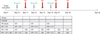
On day 10, when the average tumor size reached about 4~5 mm in diameter, each group received a different combination of treatments (Fig. 1). The following treatment methods were applied in each group; 0.5 µg/ml paclitaxel was administered through an intraperitoneal injection in mice at a dose of 5 mg/kg, RT (8.5-Gy) was performed 6 hours later and both treatments were administered again on 2 days; day 12 and day 14. The total irradiation dose was 25.5-Gy. DCs were injected intratumorally four times on days 11, 13, 15 and 18 (2×10⁶ cells/mouse for each injection in 50 µl of PBS). This injection protocol was established by Choi et al.6 with a modification described by Shin et al.20. Tumor volumes were measured using a caliper and expressed as the product of the two maximal perpendicular diameters (mm2).
Second tumor challenge experiment in the mice with complete tumor regression
A second tumor challenge experiment was performed to determine the persistence of tumor-specific immunity in mice with complete tumor regression and treated with combination therapies. Four age-matched control mice, two mice treated with RT+DCs and four mice treated with RT+Chemo+DCs were used in the second tumor challenge experiment. MCA102 cells (2×10³ cells each in 50 µl PBS) were inoculated subcutaneously in the upper left flank, the side opposite to the site of the first injection.
PKH 26 labeling of target cells
MCA102 cells (2×10⁶ cells/ml in a 2 ml total volume) were used as the target cells. These cells were transferred to polystyrene tubes and washed twice with serum-free medium before staining. Cells were then incubated for 40 minutes with freshly prepared 2 µmol/l PKH-26 (Sigma Chemical Co.) at room temperature and the staining reaction was stopped by the addition of 1 ml FBS. These cells were subsequently incubated at 37℃ for 24 hours. The splenocytes from mice with complete tumor regression after the first treatment were used as the effector cells. Labeled target cells were seeded into each well plates and incubated with the effector cells (4.8×10⁶ cells/ml) at different effector : target ratios; 1.5 : 1, 6 : 1, and 12 : 1.
Flow cytometric analysis
After the effector cells and target cells were incubated at 37℃ in 5% CO2 for 3 hours, 10 ml of 50 mg/ml of 7-AAD (Sigma Chemical Co.) solution diluted in annexin binding buffer and 5 ml of annexin V (PharMingen, San Diego, CA, USA) was added to the cell suspension for 10 minutes on ice. Samples were analyzed by a FACScan flow cytometer (Becton Dickinson Immunocytometry Systems) with an air-cooled argon-ion, 488 nm coherent beam laser. The instrument contained three optical filters, FL-1 (Annexin-FITC) at 530 nm, FL-2 (PKH 26) at 585 nm, and FL-3 (7-AAD) at 650 nm. Spectral overlap was electronically compensated using single color controls including target cells alone and cells treated or not treated with PKH 26, Annexin V or 7-AAD in separate tubes22. The percentage of dead cells (7-AAD positive, red or orange fluorescence) among PKH 26-stained target cells (green fluorescence) was calculated.
Statistical analysis
The measured value of the tumor size was expressed as the mean±standard error for each condition. ANOVA with nonparametric analysis of repeated measures was performed for comparisons between multiple groups23. A statistical analysis was performed using SPSS software ver. 12.0 (SPSS Inc., Chicago, IL, USA), with statistical significance set at p<0.05.
RESULTS
Isolation of bone marrow-derived dendritic cells and their phenotyping
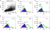
Primary DCs were obtained from syngeneic mouse bone marrow precursors in accordance with the previously described method6. The phenotypes of the DCs were verified by flow cytometry analysis (Fig. 2). Almost no expression of maturity surface markers CD40 and CD80 was detected, indicating that the DCs were in an immature state. Flow cytometry data with three other surface markers, CD11c, CD44 and I-Ad, showed the purity of the DCs to be >65%7
Significant regression of tumor size in the treated groups in contrast to the untreated control group
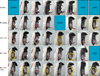
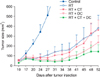
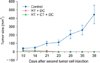
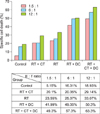
Representative photographs of the actual tumor sizes in each group on day 52 are shown in Fig. 3. Among the treated groups, the triple combination treatment group showed the highest rate of complete tumor suppression; 0% in the control group, 14.3% in the RT alone group, 12.5% in the Chemo and RT group, 28.5% in the RT and DC injection group, and 50% in the triple combination treatment group (Fig. 3). As shown in Fig. 4, the tumors grew rapidly in the untreated control mice. In contrast, the tumor growth was suppressed significantly in the treated groups (p<0.001). We observed that the triple combination therapy was more efficient than the other combination therapies in decreasing the average tumor volume. However, the statistical analysis showed no significant difference between the four treatment groups (p=0.2627). There were no side effects or any accompanying phenomenon in each group.
Persistent antitumor memory after combined treatments of radiotherapy and dendritic cell injection with/without chemotherapy
To further evaluate the memory of antitumor immunity in vivo after the combined treatments, a second challenge with the same MCA102 tumor cells was performed in the eight mice that exhibited complete tumor regression; two age-matched control mice, two mice from the RT+DC group and four mice from the RT+Chemo+DC group. We conducted the second challenge of fibrosarcoma cell injection in the RT+DC group and the RT+Chemo+DC group because these groups showed a higher antitumor effect (Fig. 4). Also, we have already shown that the tumor-free mice treated with the combined therapy of paclitaxel chemotherapy and DC injection were able to resist a repeat challenge with the same type of tumor6. As shown in Fig. 5, a very small tumor appeared around day 10 after the injection of tumor cells, but it disappeared quickly after day 14. However, the tumors grew rapidly in the control group. These results suggested that tumor-free mice treated with RT+Chemo or RT+Chemo+DC injection were highly protected against the tumor rechallenge with the same type of tumor.
Evaluation of apoptosis of MCA102 cells using an in vitro cytotoxicity assay
To examine the apoptosis of MCA102 cells after each treatment, a PKH-26 cytotoxicity assay was performed using one mouse from each group, which showed complete tumor regression. As shown in Fig. 6, the results of the flow cytometric analysis using 7-AAD and annexin-FITC showed that the CTL activity was highest in the triple combination treatment group. The splenocytes from untreated mice had a much lower CTL activity. The splenocytes from the mice treated with RT+DC also had a high level of CTL activity, but it was lesser than that in the triple combination treatment group. These results suggest that the combination of RT and DC injection can effectively induce more number of Th1 T cells and tumorspecific CTLs.
DISCUSSION
Intratumoral injection of DCs is one of the promising methods for inducing therapeutic antitumor response. However, DCs alone rarely cause curative antitumor effects as tumor microenvironment is resistant to antitumor immune response23. Therefore, another treatment is required to potentiate the effect of DC administration23. We have previously shown that the combined treatment of i.t. injection of DCs and paclitaxel chemotherapy led to tumor regression6. In this study, we evaluated the effect of RT combined with i.t. injection of DCs as an optimal combined strategy. The data showed that the RT combined with i.t. injection of DCs caused a partial regression of the tumor. However, the combination therapy of RT and i.t. injection of DCs in the second tumor challenge experiments induced acquired long-term antitumor immunity. It is possible that RT augments the antitumor efficacy of i.t. injection of DCs by modifying tumor cells to become more immunogenic. Both chemotherapy and RT induce a pro-apoptotic environment and increase the immunogenicity resulting in increased therapeutic efficacy24. The apoptosis-inducing death receptor CD95 (Apo-1/Fas) is thought to play a key role in apoptosis and is up-regulated in response to chemotherapy and RT25. Recent studies have shown that another mechanism for the immunogenic effects of chemotherapy and radiation is the toll-like receptor-4-dependent antigen processing on DCs that is dependent upon calreticulin exposure and HMGB1 release from dying tumor cells10,26. Also, these treatments induce apoptosis and necrosis of tumor cells, thereby providing DC maturation signals27.
To the best of our knowledge, this is the first report to demonstrate that i.t. injection of DCs combined with RT and chemotherapy can show enhanced therapeutic efficacy in murine fibrosarcoma. In our study, we found that the triple combination therapy of RT, paclitaxel chemotherapy and i.t. injection of immature DCs causes a decrease in the tumor volume of fibrosarcoma among various combined strategies although there was no statistically significant difference between the experimental groups. However, the triple combination immunotherapy group demonstrated a higher rate of complete regression of tumor cells and induced anti-tumor memory immune responses.
The potential explanations regarding the efficacy of the triple combination treatment are as follows: First, paclitaxel chemotherapy and RT before DC injection would decrease the tumor burden and increase the possibility of exposure of tumor antigen to DCs. Second, the high number of DCs exposed to tumor antigens would increase the activation of T-lymphocytes in the draining lymph node and thereby induce a higher degree of tumor-specific cytotoxicity. Third, i.t. injection of immature DCs would increase the chances of tumor antigen uptake and processing in vivo, as described previously by Sauter and group27. As DCs are the most effective cells that can present an antigen to MHC class I- and class II-restricted T cells28, intratumorally injected DCs could migrate from the vicinity of the tumor to the draining lymph node and induce systemic immunity29. Intratumoral administration of DCs, therefore, can increase tumor-specific CTL and Th1-type CD4 cells and decrease tumor cells30. Furthermore, a predominant role of CD8+ T cells in tumor growth inhibition upon i.t. injection of DCs has been reported31.
This study has some limitations. Firstly, we excluded mice treated with paclitaxel chemotherapy and i.t. injection of DCs from the study. Moreover, the triple combination treatment decreased the tumor volume; however, there were no statistically significant differences on comparison with the other combined strategies. Finally, a small number of mice were subjected to the secondary challenge test, which might limit our ability to reach a conclusion about the lack of association between triple combination treatment and treatment efficacy.
In conclusion, this study provides evidence supporting the potential therapeutic use of triple combination therapy in regression of tumor size, induction of systemic immune response and acquisition of persistent tumor immunity. A further research aimed at elucidating the rationale for performing more extensive clinical studies in fibrosarcoma and other types of cancer is required.
 XML Download