

 PDF
PDF ePub
ePub Citation
 Print
Print


INTRODUCTION
Epidermal nevi (EN) are hamartomatous lesions derived from epidermal components originating from pluripotent cell mutations. EN include verrucous epidermal nevus, nevus sebaceus, wooly hair nevus, and nevus comedonicus. The incidence of EN has been reported as 1 to 3 per 1,000 live newborns1,2. Their location is variable, following Blaschko lines, and reflecting embryonic migration patterns of the skin1. EN have been classified according to their predominant component; however, in some nevi, the predominant tissue may vary with the evolution of the lesion, and different areas of the same lesion may show a variety of components at the same time3. This report describes a 15-year-old girl with concurrent nevus sebaceus (NS) and nevus comedonicus (NC) with no organ involvement.
CASE REPORT
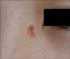
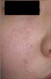
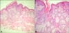
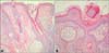
A 15-year-old girl presented with a yellowish plaque on the left medial canthus and a group of closed comedo-like papules on the right side of the cheek. She has not had any developmental problems or a history of seizures. No other family members had similar lesions. The 2 different EN lesions on the patient's face were noticed during a dermatological examination. The yellowish, asymptomatic, bean-sized plaque with a verrucous surface was noted on the left medial canthus (Fig. 1). It was present at birth. In addition, an aggregation of closed comedo-like papules and depressed pinpoint craters were found on the right side of the cheek (Fig. 2). It developed at the age of 10 years. She has also had numerous, scattered, pea-sized, hyperpigmented macules on her face for several years (Fig. 1). Her physical examination was normal except for the dermatological findings. Her neurological examination and laboratory examinations, including complete blood cell count, blood chemistry, and urine analysis, were within normal limits. Histopathological examination of the plaque on the left medial canthus showed acanthotic and papillomatous epidermal hyperplasia and large sebaceous glands located close to the epidermis (Fig. 3). Histopathological examination of the papule on the right side of the cheek revealed a dilated follicular infundibulum filled with keratin and incomplete hair follicles, consistent with the clinical diagnosis of NC (Fig. 4). She received laser treatment for NS and comedo extraction for NC in a private clinic before visiting our hospital. However, the NS of the left medial canthus recurred, and depressed craters were left with comedones on the right side of the cheek. The NS was surgically removed from the left medial canthus. The patient declined treatment of the NC and hyperpigmented macules.
DISCUSSION
EN are hamartomatous proliferations of the epithelium, including keratinocytes, sebocytes, pilosebaceous units, eccrine glands, or apocrine glands. Eighty percent of the lesions appear within the first year of life, with most of the lesions appearing by the age of 14 years. In this patient, NS appeared at birth, and NC was observed at the age of 10 years. The term epidermal nevus syndrome refers to the association of EN with extracutaneous abnormalities4. Abnormalities of the central nervous system, skeletal system, eyes, and oral cavity were reported in most cases. We did not detect any other systemic abnormality in our patient. The cutaneous features of EN depend, in part, on the predominant cell type involved, degree of cellular differentiation, location of the body part affected, and age of the patient. EN follow linear patterns known as 'the lines of Blaschko'. They seem to represent the dorsoventral migratory pathways of the neuroectoderm during embryogenesis5. The most common sites of involvement are the head and neck. In the present case, NS and NC were localized on the face following the lines of Blaschko. NS is relatively common, representing approximately onehalf of all EN6. NS is linear, hairless, yellowish-brown, waxy, and flat at birth. It becomes plaque-like with a verrucous surface owing to the hormonal changes during puberty, and usually affects the scalp, neck, and face7.
NC is a rare, sporadic epidermal nevus, characterized by an aggregation of dilated hair follicles filled with keratin plugs resembling comedones. NC was first described as an entity by Kofmann8 in 1895, and it usually has a linear or zosteriform configuration. NC appears on the face, chest, trunk, or abdomen. It may develop at any time from birth to middle age but is usually present by the second decade. NC lesions follow a noninflammatory or inflammatory course and do not resolve spontaneously. The inflammatory course may result in cyst formation with a recurrent infection, fistulae, abscesses, and scarring9. Our case showed the typical clinical findings of noninflammatory NC.
The NC in our case needed to be clinically differentiated from comedonal acne. Consistent with NC, our case showed the presence of comedones, which on extraction left a crater on the skin surface. In addition, it followed the lines of Blaschko, and it was confined to one side of the face. Besides the typical grouping of comedones, the age of onset and persistence made it easy to distinguish this condition from comedonal acne10. Histopathological examination was used to differentiate NC from comedonal acne. Unlike comedonal acne, the pilosebaceous units in NC were poorly formed. A dilated pore of Winer can sometimes be confused with NC in a histopathological examination. However, this condition is usually seen in the elderly and can be clinically differentiated.
Vidaurri-de la Cruz et al.1 evaluated 443 patients with EN. They found NS in 168 (38%) patients, and NC in 5 (1%) patients. They did not detect ≥2 EN together in their series. Köse et al.11 described a 22-year-old man with 3 different EN (NS, Becker's nevus, and NC). The patient had NS on his neck, NC on the right side of the spine, and Becker's nevus on the left shoulder. Kim and Kang12 also reported on a 21-year-old man with NC and epidermal nevus at the same site. He presented with comedo-like papules on the right buttock and thigh that had been present since infancy, and at age 14 years, he noticed the epidermal nevus lesion surrounding the comedones. Although some cases of NC associated with other EN, including verrucous linear nevus, have been reported, NC accompanying NS is very rare13. Our patient showed NC on the right side of the cheek and NS on the left medial canthus.
NS and NC are considered components in the spectrum of androgen-sensitive disorders11. It is well known that NS increases in size at puberty, revealing a correlation with the androgen hormone. Although there are no data about the androgen receptor, NC shows acneiform eruption promoted by androgen stimulation.
Facial hyperpigmented macules cannot be categorized as a particular type of EN. However, the macules of our patient were in close proximity to the NS and the NC. Melanocytic nevus is considered one of the cutaneous abnormalities in EN14. Moreover, considering that phacomatosis pigmentokeratotica is characterized by the presence of an NS and a contralateral or ipsilateral speckled lentiginous nevus, hyperpigmented macules may be associated with NS or NC15.
We report a rare case of 2 different EN, including NS and NC, with no systemic involvement.
 XML Download