

 PDF
PDF ePub
ePub Citation
 Print
Print


Dear Editor:
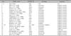
Intramuscular vascular malformations (IVMs) are rare tumors accounting for ≤1% of all hemangiomas; approximately 14% of cases are localized in the musculature of the head and neck1. This entity was previously described as cavernous hemangiomas; however, according to histological features, it is desirable to classify them into vascular malformations. The masseter muscle is the most frequently affected muscle1. IVMs of the temporalis muscle are extremely rare, with only 24 cases having been reported in the literature (Table 1)1. The actual incidence may be higher than reported, only a few cases are reported because IVM is not considered a differential diagnosis in the field of dermatology.
A 46-year-old woman presented to our department, with a history of a mass in the left temporal fossa that had been gradually increasing in size during the last 10 years. Physical examination revealed a soft, fluctuating, and nontender soft-tissue mass measuring 2×2 cm in the left temporal fossa (Fig. 1A). Computed tomography (CT) revealed a well-demarcated, heterogeneously enhanced mass measuring 2 cm in diameter (Fig. 1B). Intraoperatively, the mass was red, confined within the left temporalis muscle, and did not show any infiltration into the surrounding muscle. Histopathologialc examination revealed different sized and shaped ectatic vascular lumina that were lined with flat endothelial cells surrounded with fibrous stroma. Packed red blood cells and eosinophilic fluid were observed within the vascular lumen (Fig. 1C). Lining endothelial cells stained positive for CD31 (Fig. 1D), which is a marker of endothelial differentiation, and they were negative for SMA and D2-40. On the basis of radiological, surgical and histological findings, the tumor was diagnosed as an IVM of the temporalis muscle.
IVMs were first described by Liston as cavernous hemangiomas2, and they were classified by Allen and Enzinger3 in 1972, depending on the vessel size. Trauma and hormonal changes are considered important factors that cause ectasia of pre-existing embryonic vascular malformations4. Because they are rare and do not often exhibit any vascular signs such as pulsation or discoloration of the overlying skin, it is difficult to diagnose tumors as IVMs before radiological examination or surgical excision. Because IVMs usually present as soft, mobile and distinct tumors, differential diagnosis with neurofibromas, lipomas, dermoid cysts, and enlarged lymph nodes is necessary. Contrast-enhanced CT is useful for distinguishing IVMs from other soft-tissue tumors, for defining the size and anatomical location of the tumor, and for deciding the method of treatment. Recently, sclerotherapy has been recommended as the preferred treatment; however surgical excision remains one of the main methods of treatment4. In case the mass is observed within the temporalis muscle, careful surgical dissection during excision is important to prevent injury to the temporal branch of the facial nerve and auricular nerves. The authors totally excised the IVM through the surgical approach, and no recurrence of the tumor was observed at the 15-month follow-up visit.
Herein we report a rare case of IVM of the temporalis muscle. The authors used contrast-enhanced CT to clarify the location and characteristics of the tumor, and then they successfully excised the tumor. On the basis of this experience, we emphasize the importance of radiological examination in diagnosing benign tumors and in designing the therapeutic regimen in dermatologic clinics. When dermatologists diagnose a soft-tissue mass, IVM should be included in the differential diagnosis.

 XML Download