

 PDF
PDF ePub
ePub Citation
 Print
Print


Dear Editor:
Dermatologists instinctively consider syphilid when viewing discrete macular or papular lesions on the palms, which may be an important clue in distinguishing syphilid from other disorders. The lack of clinical analysis on syphilid eruptions on the palm prompted us to investigate specific clues in differentiating syphilid from other disorders. We analyzed the characteristics of patients with syphilid (n=34) or syphilid-like eruptions (n=39) on the palms through a retrospective comparison, by using clinical photos and medical records from Pusan National University Hospital (PNUH) between January 2002 and July 2011. The study was approved by the ethics committee of PNUH, and voluntary informed consent was obtained in written form from all of the participants.
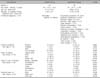
The syphilid-like group consisted of patients with discrete macular or papular lesions on the palms, mimicking typical syphilid but with negative treponemal and nontreponemal test. The definition for the syphilid-like group was based on the description in Fitzpatrick's textbook: "Palmoplantar lesions may be macular or papular, discrete or diffuse, and non-scaling, slightly scaly, or hyperkeratotic." 1 The syphilid-like group (n=39) included 10 (25.6%) patients with eczematous dermatitis, 8 (20.5%) with erythema multiforme, 6 (15.4%) with psoriasis, 3 (7.7%) with pityriasis rubra pilaris, 2 (5.1%) with drug eruption, and 10 (25.6%) with other skin conditions (Table 1).
Concerning palmar lesions, the differences between the two groups were analyzed in terms of symptoms, color, bilaterality, and scale types. For comparative analysis, cross-tabulation and independent t-test were used with PASW ver. 18.0 for Windows (IBM Co., Armonk, NY, USA). A p-value of ≤0.05 was set as the level of statistical significance.
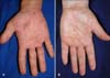
The patients in the syphilid-like group (30 of 39, 76.9%) were more symptomatic than those in the syphilid group (16 of 34, 47.1%) (p=0.008). Concerning the color of lesions, more dusky red lesions were found in the syphilid group than in the syphilid-like group (35.3% vs. 10.3%, p<0.05). Biette's collarette was present in 76.4% (26 of 34) of patients in the syphilid group and 23.1% (9 of 39) of patients in the syphilid-like group, with a statistically significant difference. In the syphilid-like group, scaling was not observed in 17 patients (43.6%) and Biette's collarette had the highest percentage (23.1%, 9 of 39) among the scale types. In terms of bilaterality, there was no significant difference between the syphilid group (94.1%, 32 of 34) and the syphilid-like group (87.2%, 34 of 39). The typical lesion of each group is shown in Fig. 1.
The results of this study confirmed that palmar syphilid could have various colors and scale types. To summarize the most common and statistically significant characteristics in the syphilid group, asymptomatic dusky red macules or papules having Biette's collarette on both palms can be considered as the typical presentation. However, about half of the patients in the syphilid group were symptomatic and only 35.3% of them showed dusky red lesions. In addition, the dusky red color was observed in about 10% and atypical scales in one-fourth of the syphilid-like group. This study is noteworthy because it provides the standard characterizations for typical and atypical lesions through a comparison with the syphilid-like group.
This study has some limitations. The number of patients was not sufficient to make an accurate statistical analysis, and the syphilid-like group consisted of patients with various diseases. However, this study is the first to compare and analyze syphilid and syphilid-like eruptions on the palms and will help deepen the clinical understanding of these conditions.
 XML Download