

 PDF
PDF ePub
ePub Citation
 Print
Print


Dear Editor:
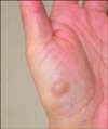
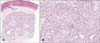
Nodular hidradenoma (NH) is a benign, rare adnexal neoplasm. NH usually occurs on the scalp, trunk, and proximal extremities, and rarely on the hands and feet1. The subdivision of NH into two groups was recently suggested, as follows: those with eccrine differentiation (known as poroid hidradenoma [PH]) and those with apocrine differentiation (known as clear cell hidradenoma [CCH])2,3. To our knowledge, there have been no previous reports of CCH on the volar surface of the hands and feet. Herein, we report a case of CCH on the palm. A 51-year-old Korean woman without any underlying diseases presented with a brownish nodule on the thenar area of her left palm (Fig. 1). Histological examination revealed a circumscribed dermal neoplasm comprising several aggregations of neoplastic cells and cystic areas (Fig. 2A). Epidermal connection was observed. Most of the tumor cells were polygonal cells with eosinophilic cytoplasm, and few clusters of clear cells were observed (Fig. 2B). Apocrine differentiation was not observed. Immunohistochemical analysis revealed positive expression of cytokeratin (CK) 5/6 in the neoplastic cells. Carcinoembryonic antigen and epithelial membrane antigen (EMA) were partially expressed in the ductal structures and polygonal cells. The clear cells were negative for both EMA and androgen receptor (AR). Histologically, NH mostly is a dermal neoplasm presenting as a well-circumscribed lobulated mass. Occasionally, an epidermal connection can be identified4. Tubular or ductal structures with luminal and cystic spaces can be observed. The intervening stroma of NH shows various patterns including sclerotic, vascularized, and myxoid patterns. CCH is composed of polygonal cells and clear cells. Clear cells usually account for 5% to 30% of the total tumor cell mass3. PH is composed of poroid cells and cuticular cells. In this case, some clear cells were observed between the predominant polygonal cells, and thus, we diagnosed this case as CCH (Fig. 2B). Although the origin of CCH is controversial, it has been considered that it usually arises from folliculo-sebaceous-apocrine units; however, it can also originate from eccrine units2. To determine the origin of this tumor, we immunostained the specimens with monoclonal AR antibodies; however, the clear cells stained negative for AR antibodies. The positive staining for CK5/6 and the connection with the acrosyringium in this case suggest that this tumor originated from the basal layer5.
Owing to the site of the tumor in the area without folliculo-sebaceous-apocrine units and the connection with the acrosyringium, we believe that this case of CCH originated from the acrosyringium of the eccrine gland.
 XML Download