

 PDF
PDF ePub
ePub Citation
 Print
Print


INTRODUCTION
The nose involving the ala and perialar area presents a reconstructive challenge after tumor excision because of its complex architecture and cosmetic subunits1,2. Although there are many options for repairing deep alar defects, including two-stage interpolated cheek flap and paramedian forehead flap, none of them are ideal3,4,5,6,7,8,9,10,11. Lateral alar wounds of full thickness usually cause various problems in nasal function and aesthetic balance, and often require multistep repair techniques such as inner lining replacement, cartilage batten graft support, and outer skin coverage. Spear et al.12 in 1987 described a single operative procedure, the reverse nasolabial flap, for reconstructing difficult full-thickness, lateral alar defects. We successfully performed this one-stage repair, with minor modifications, in a patient with basal cell carcinoma showing a full-thickness alar defect spread over three cosmetic subunits, the ala-cheek-lip junction. In addition, a free cartilage graft from the ear was used concurrently to keep the airway open.
CASE REPORT
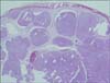
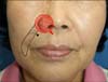
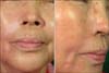
A 65-year-old woman presented with a chronic asymptomatic nasal lesion. Physical examination showed a small, elevated nodule on the right lateral ala. The nodule was skin-colored, 1.0×1.5 cm in size, round and firm, and had telangiectasia on the surface (Fig. 1). Skin biopsy revealed micronodular basal cell carcinoma (Fig. 2). We decided to perform surgery and explained to the patient the various surgical methods available, including twostage procedures, but she preferred a single procedure. Thus, we chose the reverse nasolabial flap. Initially, the lesion was removed with a surgical margin of 4 mm; however, the specimen was marginally positive in the frozen pathology evaluation, and three more examinations were required to extirpate the tumor. The resultant defect was loss of the lateral ala involving the alar rim, crease, and the adjacent cheek, leaving a 'through-and-through' type of full-thickness defect. The flap was designed along the nasolabial fold and was deepithelialized in the proximal one-third portion with a No. 11 blade (Fig. 3). The flap was elevated in the subcutaneous plane and was turned over upward and medially like a page of a book. The proximal flap was sutured to the lining side of the defect. The remaining portion of the flap was then folded onto itself above the grafted cartilage brace for the outer coverage. The free cartilage graft, 1.5×0.5 cm in size and rectangular in shape, was harvested from the conchal bowl and fastened medially and laterally to the defect with Vicryl 5-0 sutures. After the flap was trimmed and sutured into an appropriate position, the donor sites of the face and ear were closed primarily. During the follow-up period of 3 years, no complications were observed in function and aesthetics, and the patient has remained in a disease-free state (Fig. 4).
DISCUSSION
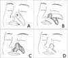
The Spear flap provides both inner lining and outer coverage by folding itself like the turning of a book page, and is well suited for full-thickness lateral alar defects (Fig. 5)4,13. We modified this flap to fill a very deep cheek defect. Before the first turnover of the flap, we deepithelialized its proximal portion in advance. In designing the flap, the base was placed more inferiorly and medially than standard nasolabial flaps, and placed as near to the site of the original ala-cheek-lip junction as possible so as not to displace the alar base laterally. The medial border of the flap corresponded to the nasolabial fold and the lateral border was placed in parallel to the fold. It is important that the flap is wider than the nasal defect7.
The choice of the repair method depends on the presence of the alar rim and groove after tumor removal. If they are preserved or if the defect is confined to one or two subunits, e.g., the ala-lip junction, reconstruction is rather easy and excellent cosmetic result could be expected3,8.
Some single-stage procedures for repairing ala-perialar defects include the use of shark island pedicle flap9, nasolabial flap10, tunneled transposition flap7,11, composite cartilage and skin graft, and combinations of these techniques. Tunneled transposition flap and the auricular composite graft are strictly indicated for defects located entirely on the ala. Shark island pedicle flap resembles a shark with a snout and ventral mouth, and was developed for a combined ala-perialar defect with an intact alar rim. Zitelli's nasolabial flap as a single-stage procedure also has certain limitations. It is not feasible for defects that extend onto the cheek and has well-recognized disadvantages such as blunting of the nasofacial angle and causing trapdoor deformity.
Usually, the turnover flap does not need a cartilage support5,12. However, our patient showed partial nostril collapse when the flap was folded secondarily, which needed a free cartilage support graft such as alar batten. The cartilage graft, consisting of cartilage with overlying perichondrium and harvested from the ear, was gently scored and sewn into place, bent outward to avoid alar collapse and to recover the alar contour12. This flap has many advantages4,5,6,7,11,12. First, it does not need several staged operations for replacing the inner nasal mucosa, structural support, and outer skin coverage. Second, the flap is based on a rich vascular supply from the angular artery, which eliminates the possibility of flap necrosis through multiple turnovers. As mentioned above, because this single-stage flap has a tendency to displace the alar base laterally, the alar base should be rotated more medially and inferiorly. Such a rich vascular supply can minimize flap necrosis during this manipulation. Third, primary closure of the donor site yields an acceptable scar mimicking the natural nasolabial fold. One disadvantage is that the ala is lined internally by skin, not by mucosa; however, this is rarely a problem with daily lubricant application5. In conclusion, the reverse nasolabial flap with a cartilage graft can offer a single-stage repair of difficult alar defects, especially lateral ala involving a complete loss of the alar rim and crease.

 XML Download