

 PDF
PDF ePub
ePub Citation
 Print
Print


INTRODUCTION
Pterygium inversum unguis (PIU) is rare nail abnormality characterized by the adherence of the distal portion of the nail bed to the ventral surface of the nail plate, resulting in a subungual extension of the hyponychium and obliteration of the distal groove1,2. The term 'pterygium inversum unguis' was first described in 1973 by Caputo and Prandi3, and its cause is not well understood. This rare condition may be congenital or acquired. The acquired forms, which may be idiopathic or secondary to systemic connective tissue diseases or other causes, have been reported in a few patients. Herein, we report a rare case of PIU of all 10 fingernails, which is the first report of its kind in the Korean dermatologic literature.
CASE REPORT
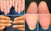
A 22-year-old healthy man presented with aberrant nails on all 10 fingers, a condition that he had since childhood. He had relatively long fingernails because he did not clip his nails very often owing to pain, discomfort, and easy bleeding that resulted from nail clipping. He had no other symptoms, history of systemic disease, previous trauma, nail disease, or a family history of a similar abnormality. Physical examination of the fingers showed overgrowth of the hyponychium attached to the ventral surface of the free edge of the distal part of the nail plate, and marked subungual thickening owing to keratosis, with loss of the nail grooves of all 10 fingers (Fig. 1). His toenails showed a normal appearance. A punch biopsy specimen from the center of the right index fingernail did not show any specific findings on histopathological examination. We could not conduct further evaluation or administer treatment because the patient was staying abroad.
DISCUSSION
The first case of PIU was described in 1973 in a woman who developed ventral pterygium on multiple fingers of both hands without any apparent causes3. Initially, the authors summarized this abnormality to be an acquired, non-familial, gradually developing, symmetrical, pathologic entity that did not alter the nail plate or periungual soft tissues.
From the time of the first description, 37 cases have been reported in the English literature (Table 1)1,2,3,4,5,6,7,8,9,10,11,12,13. Among the 37 reported cases of PIU, 34 were acquired and 4 were congenital; 26 patients were women and 12 were men. Three patients had a positive familial history. In 12 cases, there were no associated abnormalities, whereas associations with different diseases were reported in the other 25 cases. In most patients, PIU was associated with connective tissue diseases such as systemic sclerosis or lupus erythematosus5,8. Other postulated associations were acrylate allergy, acrylic nail uses among women11, use of nail fortifiers12, stroke2,7, neurofibromatosis5, and leprosy13. Most patients showed PIU lesions on both hands, on either all or some of the fingernails. The toenails are rarely affected, with only 3 reported cases of PIU involving the toes1,7,9. The patient in this case was a 22-year-old man with extra skin at the base of all 10 of his fingernails since childhood. This case is an acquired idiopathic form of PIU affecting all the 10 fingernails without involvement of the toenails.
The cause of PIU is unknown. Congenital PIU is thought to be caused by an early defect in the development of the fetal groove and ridge. This abnormal embryonic onychogenesis causes mechanical stretching of the hyponychium, which consequently undergoes distal migration through its attachment to the growing nail plate4. Caputo and Prandi3 suggested that idiopathic forms of PIU might be due to the distal extension of the zone of the nail bed that normally contributes to the formation of the nail plate. Patterson5 reported that secondary PIU is the consequence of abnormal distal circulation. Ischemia and altered blood flow result in recurrent ulceration and scarring, which leads to hyperkeratosis of the stratum corneum and pterygium formation. This was hypothesized as the possible pathologic mechanism for pterygium formation in patients with leprosy13 or stroke2,7. However, all these pathophysiologic hypotheses are speculative.
Three cases of PIU have been histopathologically investigated1,2,10. Histological examination revealed a marked hyperkeratotic stratum corneum that extended and firmly attached to the undersurface of the nail plate with normal nail bed vasculature. Vadmal et al.2 concluded that vascular and neurosensory disturbances might have contributed to reactive hyperkeratosis in their patient with stroke. Recently, Oiso et al.10 observed a marked, eosinophilic, keratinized substance with nucleated corneocytes attached to the distal and ventral nail plate, and a whorled keratinized substance in the horny layer of the fingertip. They suggested that the keratinized structure was derived from the nail isthmus, and that PIU was possibly caused by aberrantly regulated keratinization in the isthmus.
The management of PIU is not well defined; different treatments including the use of keratolytics or topical steroids as well as surgical excision with electrocautery are reported to be ineffective4,7,9. The most effective strategy is the treatment of the underlying cause of PIU11. Patients with PIU need to be evaluated to rule out associated rheumatologic diseases, and require long term follow-up examinations to detect the development of connective tissue diseases such as scleroderma. In our case, unfortunately, the patient could not receive any treatment or further long term follow-up because he was living abroad. Future investigations involving a large number of cases will result in a more accurate classification as well as clarification of the pathogenesis of PIU, and the determination of an effective form of therapy.
 XML Download