

 PDF
PDF ePub
ePub Citation
 Print
Print


INTRODUCTION
Irritant contact dermatitis (ICD) is a common occupational skin condition and a risk factor for allergic contact dermatitis1. A study conducted in Germany reported that 32% of surveyed junior hairdressers experienced chronic irritant contact dermatitis (CICD) during their first year in the occupation, with repetitive exposure to the offended materials diminishing the prevalence of clinical symptoms by 90%2. This long course of diminished non-specific responses to chronic repetitive exposure is termed the hardening phenomenon or accommodation3-5. It describes the condition when ICD disappears despite continued exposure to the irritant and avoidance of CICD3-6.
The mechanism of this skin hardening remains elusive. The degree and duration of stimulation required to induce skin hardening are unclear. Further studies are needed to prevent CICD and to detect individuals sensitive to irritants.
One theory that can explain the decreased reactivity of the hardened skin to irritants is altered barrier function. Patients with skin conditions like atopic dermatitis are more prone to CICD, suggesting that the skin barrier is important in the pathogenesis of CICD7. Altered lipid composition of the stratum corneum layer may contribute to skin hardening. Ceramides, which are lipids that function in the skin barrier, are key role in the protection mechanism against irritants8,9. Heinemann et al.10 reported that the amount of ceramide 1 was increased in response to 3-week stimulation with 0.5% sodium lauryl sulfate (SLS) solution. Whether ceramide contributed to the hardening phenomenon was not conclusively established. The aim of this study was to compare the effects of two concentrations of SLS on the induction of the hardening phenomenon by assessing changes in transepidermal water loss (TEWL) in healthy Korean volunteers, and to measure the levels of ceramides of the horny layer in the hardened and non-hardened skin.
MATERIALS AND METHODS
Subjects and materials
The study included 41 volunteers (24 males, 17 females) free from known chronic diseases who visited the Department of Dermatology, Kangnam Sacred Heart Hospital, Hallym University. The average age of the patients was 25.8 years (range, 22 to 35 years). This study was approved by the Institutional Review Board of Hallym University Kangnam Sacred Heart Hospital, and informed consent was obtained. Subjects did not have a past or familial history of atopic dermatitis, or any other chronic skin disease. During the study, all subjects were allowed to bathe as usual and were instructed to avoid direct application of detergents, moisturizers, emollients or topical medications to their forearms. From 2 weeks before beginning the study, all subjects were prohibited from using any topical or systemic medications.
Measurement and instrumentation
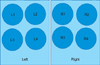
Each patient's skin was marked with four 20-mm-diameter separated circles in the proximal one-third of both forearms using an oil-based pen (left, L1~L4; right, R1~R4) (Fig. 1). Before the test, TEWL and erythema index (E-index) were measured in all eight sites of both forearms. TEWL and E-index were measured under conditions without air flow and direct sunlight, 30 minutes after exposure of the sites to the irritants. TEWL was measured using a Tewameter 210 probe (Courage+Khazaka Electronic Co., Kölon, Germany) and E-index using a Dermaspectrometer (Cortex Technology, Hadsund, Denmark) at room temperature of 20℃±2℃ and 35% to 53% relative humidity at the same time in the afternoon. TEWL was adjusted at baseline by subtracting one standard deviation (SD) of normal for each parameter using '10' as the baseline adjustment for TEWL (mean+1 SD).
Irritant exposure and irritant contact dermatitis induction (Phase 1)
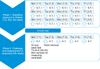
The study protocol consisted of 3 weeks of irritant exposure and ICD induction (Phase 1), 3 weeks of rest and 1 week of challenge (Phase 2) (Fig. 2). From site L1, a skin sample was collected to measure the amount of ceramides in the horny layer. After 800 µl of distilled water was applied with a micropipette L2 and R2 and dried, 0.1% SLS was applied to L3 and R3, and 2% SLS solution to L4 and R4 with a 20-mm-diameter plastic ring. Each site was washed with running water. In the same way, each site (L2~L4, R2~R4) was stimulated repetitively with distilled water, 0.1% SLS solution and 2% SLS solution for 3 weeks, 5 times per week (Monday to Friday). TEWL and E-index were measured before application and after each of 15 applications per subject (Phase 1). After that, patients had 3 weeks of rest.
Challenge (Phase 2)
Immediately after the aforementioned sites (L2~L4, R2~R4) were challenged by the same procedures for 1 week, TEWL and E-index were measured on sites L2~L4 and R2~R4 as well as the control site (site R1). Skin samples were collected from sites L2~L4 to measure ceramide levels. The 24-hour patch test was carried out using 60 µl of 1% SLS on sites R2~R4 and R1 with a large Finn chamber (inner diameter 1.2 cm; Epitest Ltd., Hyrlä, Finland) and filter paper. TEWL and E-index was measured 30 minutes after removal of the patch for the next 3 consecutive days (Wednesday, Thursday, and Friday).
Measurement of ceramides in the horny layer
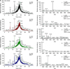
Ceramides were measured as previously described11. Briefly, the stratum corneum samples were collected from participants. To collect these samples, each site was gently swabbed with ethanol. After 1 drop of Aron alpha cyanoacrylate resin (Toagosei, Tokyo, Japan) was put on a glass slide, the sample was placed in the resin and compressed with weak pressure for 1 minute. The horny layer that had stuck to the glass slide was raked up with a scalpel blade and put onto a glass bottle. A 95:5 mixture of hexane and ethanol (5 ml) was added to the glass bottle to melt the horny layer, which was and then ultrasonicated (Sonic & Materials, Newtown, CT, USA) for 20 minutes. The contents were removed using a syringe. After membrane filtration (HV 0.45 µm, MIllex; Millipore, Billerica, MA, USA), the solution was put onto a new glass bottle. Only horny layer lipid remained in the filtrates. The solvent was evaporated with nitrogen gas and then the extract was kept at -18℃. The extract was dissolved in 1 ml of methanol, transferred to a 1.5 ml microtube and centrifuged to obtain the supernatant used for high performance liquid chromatography (HPLC) analysis. HPLC analysis was performed using a Finnigan Surveyor system (Thermo Fisher Scientific, Waltham, MA, USA) and Unison UK-C8 analysis column (250×3 mm) at a flow rate of 0.4 ml/min. The mobile phase was assessed with distilled water and methanol from a gradient of 15% B to 85% B over 23 minutes. The detector was a LCQ Advantage Max mass spectrography system (Thermo Fisher Scientific). For the improvement of mass ionization, 0.1% ammonium hydroxide, as the shield liquid, was injected at a velocity of 10 µl/min using a L-6200 pump (Hitachi, Tokyo, Japan). Electrospray ionization was used for positive ion mode analysis under the following conditions: sheath gas flow, 280 arb; spray voltage, 5 kV; capillary temperature, 280℃; and capillary voltage, 3 V. Quantities of stratum corneum ceramides were measured by HPLC-mass spectrometry (HPLC-MS). In the graph of HPLC-MS for straum corneum lipids, the sum of the peak values of lipids with molecular weights of 506, 559 and 563 were regarded as the total quantity of ceramides. The peak value of lipids with a molecular weight of 563 was evaluated separately (Fig. 3).
Statistical analyses
All statistical analyses were performed using SPSS 18.0.0 (PASW Statistics; IBM Co., Armonk, NY, USA). TEWL, E-index and the amount of horny layer ceramides were measured in each skin site at each experimental time point after repetitive application of SLS solution and distilled water. Repeated measures of ANOVA with subsequent post hoc pair-wise comparison were applied to analyze the changes of TEWL and the E-index in the different concentration groups during Phase 1 and 2. One-way analysis of variance was used to find the difference of subjects showing decreased TEWL after patch test in the three groups during Phase 2. The Scheffe method was used for multiple comparisons. The relationship between ceramides amount and hardening phenomenon was analyzed by Fisher's exact test. Differences were considered to be significant at p<0.05.
RESULTS
Changes in transepidermal water loss and erythema index in irritant contact dermatitis induction phase (Phase 1)
1) Changes in transepidermal water loss and erythema index
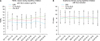
During Phase 1, TEWL was measured 30 minutes after application of 0.1% and 2% SLS. Repeated measures ANOVA revealed a significant difference in TEWL with time (p=0.004) and between the three different skin sites (control, 0.1% and 2% SLS skin sites; p=0.022). TEWL was 20.80% higher at the sites receiving 0.1% SLS compared with the control sites (10.42 vs. 8.62 g/m2h) and 33.74% higher at the 2% SLS sites compared with the control sites (11.53 vs. 8.62 g/m2h). Multiple comparison analysis revealed significant differences between control and 0.1% SLS, and control and 2% SLS (p<0.001), but not between 0.1% and 2% SLS (p=0.381). There was no intersexual variation in TEWL (Fig. 4A).
The E-index measured in the same manner was not significantly different between the 0.1% SLS, 2% SLS and control sites (p=0.432). The E-index was 1.74% higher at the 0.1% SLS sites than at the control sites (8.73 vs. 8.58, p=0.991) and 6.61% lower (8.05 to 8.58, p=0.923) at the 2% SLS sites than at the control sites. There was no intersexual variation in E-index (Fig. 4B).
Changes in transepidermal water loss and erythema index after challenge (Phase 2)
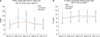
After the 24-hour patch test using 1% SLS, TEWL were measured at sites R3 and R4 stimulated by 0.1% and 2% SLS during Phase 2 (Fig. 5A). There were no significant differences in TEWL between the two sites (p=0.967). However, TEWL at site R2 stimulated by distilled water was 8.45±2.46 g/m2h before the patch test on day 43 (D43), increased to 18.49±7.84 g/m2h on D44 and to 15.50±6.26 g/m2h on D45 after removal of the patch. It subsequently decreased to 11.60±3.25 g/m2h on D46 and to 10.80±2.97 g/m2h on D47. The values of R2 were higher than both R3 and R4 on D44 (p=0.150 and 0.053) and on the total 5 days of Phase 2 (p=0.905 and 0.781) without statistical significance. After the patch test, 30 of 41 subjects showed an increase in TEWL at site R2 (distilled water). The remaining 11 subjects displayed a decrease. In addition, 25 subjects showed an increase in TEWL at site R3 (0.1% SLS solution) on D44, which decreased over time. The remaining 16 subjects showed a decrease in TEWL after the removal of the patch. Of all 41 subjects, 18 showed an increase in TEWL at site R4 (2% SLS solution) and 23 showed a decrease. There was significant difference between control (R2) and irritated sites (R4) (p=0.021), but none between R2 and R3 (p=0.521) or between R3 and R4 (p=0.280). There was no intersexual variation in TEWL. The study population was divided into two groups depending on the difference in TEWL between prestimulated and non-stimulated sites after the 24-hour patch test on D44 (7-2): those with evidence of skin hardening (negative difference) and those without (no difference or positive difference). Hardening phenomenon occurred in 24 of 41 volunteers at day 44: six at the sites exposed to distilled water, 13 at the sites exposed to 0.1% SLS and 24 at the sites exposed to 2% SLS solution. Skin hardening occurred at the sites exposed to higher concentrations of SLS solution (p<0.05).
There were no significant differences in E-index between the 0.1% SLS, 2% SLS and distilled water sites after the 24-hour patch test with 1% SLS solution. E-index was 0.11% higher at the 0.1% SLS sites and 5.84% lower at the 2% SLS sites than controls, but the differences were not statistically significant (p=0.759). There was no intersexual variation in E-index (Fig. 5B).
Changes in ceramide levels in the stratum corneum after repeated stimulation with both concentrations of sodium lauryl sulfate
Thirty exposure sites (R2, R3 and R4 of 10 volunteers) were compared to 10 sites of non-stimulated areas (R1 of 10 volunteers) as controls in 10 among 41 volunteers who agreed to collect samples from their skin (the remaining 31 participants refused to provide skin samples). Skin hardening occurred in 13 sites in six of the 10 participants on D44: three at the sites exposed to distilled water, four at the sites exposed to 0.1% SLS and six at the sites exposed to 2% SLS. Comparing the baseline quantity of stratum corneum ceramides between the sites with skin hardening (n=13) and those without (n=17) demonstrated that the total basal ceramide was slightly higher in the sites with the skin hardening than in those without, but the difference was not statistically significant (4.31±3.92×10⁸ optical density [O.D.] at the sites with skin hardening vs. 3.56±3.73×10⁸ O.D. at the sites without). There was an increase in ceramides levels at three sites and a decrease at 10 sites of the 13 sites with skin hardening. In the sites without skin hardening, ceramides levels were increased at three sites and decreased at 14 sites of the 17 sites. The mean delta value of ceramide levels determined by optical density was not significantly different between the sites with skin hardening (4.41±4.61×10⁸) and those without (4.13±4.65×10⁸) (p=0.531). There was no intersexual variation in ceramide levels.
DISCUSSION
The mechanism of skin hardening after chronic irritation remains elusive, and the degree and duration of stimulation required to induce skin hardening are contentious. This study attempted to reproduce an experimental hardening model and to establish a standard duration for the application and concentration of irritants. Skin hardening occurred more frequently at the higher concentration of SLS (2%) than at the lower concentration (0.1%). This pattern is similar to earlier studies, which reported that higher SLS concentrations more easily induce accommodation in the skin12. However, in our study, skin hardening occurred in 43.3% of subjects 3 weeks after daily application of SLS solution, which was more frequent than previously reported. This difference may be because we applied irritants for 10 minutes a day, while other investigators used the 24-hour patch test. Also, our results differ from those of a previous study, which showed that concentrations of SLS solution are not related to the incidence of skin hardening12. Branco et al.12 reported that relatively long (15-week) repetitive applications of 0.025% and 0.075% SLS to the skin did not produce skin hardening. However, Widmer et al.13 showed that after 12-week repetitive applications of 0.5% and 2% SLS, daily 1-hour applications of 0.5% SLS increased TEWL during the first 2 weeks and decreased it in the third week. In their study, as compared to the control sites (distilled water), the 24-hour patch test with 2% SLS at the SLS-exposed skin showed skin hardening in weeks 3, 6 and 9. Skin hardening typically changes appreciably in the sixth week. Heinemann et al.10 conducted a 12-week study with 0.5% and 1% SLS and confirmed the results of the study by Widmer et al.13. However, they also showed different results that skin hardening frequently occurred in the third week, did not occur in up to eighth week and occurred again in the ninth week.
Our study assessed two different SLS concentrations, by repetitive 3-week stimulation with a low concentration (0.1%) of SLS and a high concentration (2%) of SLS. After a 3-week rest, the 24-hour patch test with 1% SLS was performed to compare skin hardening between the sites exposed to the two concentrations and untreated (control) sites. The control sites showed a significant elevation in TEWL, whereas the sites exposed to both SLS solutions did not display elevation or decline in TEWL, suggesting skin hardening. Of the 41 subjects in our study, 16 (39%) showed skin hardening after exposure to 0.1% SLS and 23 (56.1%) showed after exposure to 2% SLS. The data suggest that repetitive stimulation with more concentrated SLS can more easily trigger skin hardening. However, both high and low concentrations of SLS did not induce skin hardening in some subjects, likely due to individual differences of susceptibility to inflammatory responses14. Three-week additional duration of stimulation with a low concentration of SLS may cause frequent skin hardening in subjects and repeated patch tests after 3 weeks might increase the incidence of skin hardening in those who had not shown skin hardening10.
The undamaged, normal horny layer is one of the most significant functional structures in the skin barrier. Keratinocytes synthesize lipids, which accumulate in lamellar granules, forming a layer containing ceramides, cholesterol and fatty acids through myriad enzymatic reactions. Of these lipids, ceramides have been centrally implicated in skin barrier function8,9. The symptoms due to damaged skin barrier function are improved by application of external ceramides15. Skin ceramides are reduced in atopic dermatitis.
The mechanisms and pathways involved in hardening phenomenon have not been fully elucidated. The most acceptable theory, the physical change in the skin barrier, can be explained by two aspects. The hyperkeratotic stratum corneum and thicker layer of stratum granulosum in hardened skin reduces the penetration of irritants16,17. In addition, decreased inflammatory responses to irritants is due to changes in the lipid components of the horny layer. The horny layer contains intercorneocyte lipis, consisting of ceramides, cholesterol, fatty acids and other lipids. Among them, ceramides are the most important in skin barrier function8,18, and its alteration causes changes in inflammatory responses. Since irritants increase lipid synthesis in the skin and cholesterol synthesis plays a major role in the early stages of ICD, increases in ceramides have been implicated in CICD19. The skin with a hardening phenomenon shows an increase in ceramide I, but not cholesterol and fatty acids, whereas the skin with no hardening phenomenon shows a decrease in ceramide I or an increase in fatty acids, suggesting that ceramide I synthesis plays an important role in skin hardening10,20,21. Our study was designed to evaluate the correlation of overall ceramide level on the effect of irritants in the stratum corneum and concentrations of irritants on skin hardening. The changes in ceramide levels were not significantly higher in the hardened skin than in the non-hardened skin.
Instead of ceramide, other factors, such as filaggrin or proinflammatory cytokines, might be more important for the hardening phenomenon7,22. As well, changes in the skin barrier may due to reactive hyperkeratosis. Reactive hyperkeratosis seen in the horny layer of the skin globally thickens the skin barrier and reduces inflammatory responses16. In this study, E-index was not significantly different between the hardened and non-hardened skin.
Recent studies have emphasized the decline in inflammatory responses caused by the changed composition of lipids in the skin as the mechanism of skin hardening, and TEWL has been widely used to objectively evaluate the degree of skin hardening. Our study assessed TEWL and E-index for skin barrier function and inflammatory responses to stimulation with irritants. TEWL showed a significant correlation between repetitive exposure and skin hardening, but E-index did not. Based on this result, it is thought that the change in skin barrier function by the alteration of the lipid composition may be more important in inducing skin hardening than overall inflammatory response. Further studies of factors other than ceramides are needed to explain why some participants show skin hardening without an increase in ceramide levels.
Limitations to this study include the application of a single irritant and the short duration of exposure to the irritant.
In conclusion, repetitive stimulation with higher concentration of SLS (2%) can more easily trigger skin hardening than that with lower concentrations (0.1%) in healthy Korean volunteers. The changes in ceramide levels were not higher in the stratum corneum of hardened skin than in the non-hardened skin. There seems to be another mechanism of hardening phenomenon other than ceramide in the stratum corneum.
 XML Download