

 PDF
PDF ePub
ePub Citation
 Print
Print


INTRODUCTION
Dermatophytes are pathogenic, keratinophilic fungi that invade the stratum corneum of epidermis, hair, fingernails, and toenails of animals. Throughout the world, 43 species of dermatophyte fungi have been identified. Among these, 20 are known to infect humans and other animals. Trichophyton mentagrophytes var. mentagrophytes, T. mentagrophytes var. interdigitale, T. tonsurans, T. verrucosum, T. schönleinii, T. rubrum, Microsporum canis, M. gypseum, and Epidermophyton floccosum have been reported in South Korea. Of the nine species, T. rubrum is the most abundant and accounts for 85% to 90% of all reported cases of infection. Accurate differentiation of dermatophyte subspecies of T. rubrum, T. tonsurans, and T. mentagrophytes var. interdigitale is, therefore, of clinical significance.
Dermatophytes can be differentiated by the variation in the colors and shapes of colonies when observed under the microscope and in culture. Nevertheless, differentiation of dermatophytes by these methods remains difficult. Therefore, other molecular techniques need to be considered. A variety of methods, including mitochondrial DNA restriction fragment length polymorphism (RFLP) pattern and chitin synthetase 1 nucleotide sequence analysis, have been used for simple, fast, and accurate identification of dermatophytes. Recently, the nucleotide sequences of internal transcribed spacer (ITS) regions that represent organism diversity have been analyzed.
However, the ITS sequences of T. mentagrophytes, T. tonsurans, T. rubrum and M. gypseum are very similar, making differentiation using this method difficult. Moreover, this method requires the utilization of a variety of restriction enzymes1,2,3,4.
It has recently been reported that proteins from various organisms are damaged upon the activation of proteolytic enzymes, such as metalloproteinase-1, upon exposure to ultraviolet light or high temperatures. It was found that during the recovery of the damaged DNA sequences, slight alterations occur in the sequences. These alterations often result in speciation. Recently, transgenic research using the proteolytic enzyme metalloproteinase-1 has gained popularity in the area of plant molecular biology. This method can also be applied to the study differentiation in dermatophytes and may prove to be more accurate than other known molecular methods.
In this study, common pathogenic species of dermatophytes, T. rubrum, T. mentagrophytes var. mentagrophytes, T. tonsurans, T. mentagrophytes var. interdigitale, M. canis and M. gypseum were isolated and identified using the polymerase chain reaction (PCR)-RFLP technique. The gene coding for metalloproteinase-1 (MEP1) was analyzed for polymorphisms upon restriction digestion with BsrDI.
MATERIALS AND METHODS
Bacterial species
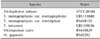
The standard species of T. rubrum, T. mentagrophytes var. mentagrophytes, T. mentagrophytes var. interdigitale, T. tonsurans, M. canis and M. gypseum were spread evenly on Mycosel agar culture medium (Papaic digest of soybean meal 10 g, dextrose 10 g, cycloheximide 0.4 g, chloramphenicol 0.05 g and bactoagar 15.5 g), sterilized at 121℃ for 15 minutes, and incubated at 34℃ for 14 days (Table 1).
Methods
1) DNA extraction
To collect the samples with roots, a 0.5×0.5 cm×1 to 2 mm block of Mycosel agar with colonies was placed in a 1.5-ml eppendorf tube (e-tube) and centrifuged at 13,570×g at 4℃ for 10 minutes. The samples were resuspended in 200 µl of Lysis buffer (100 mM Tris-HCl pH 9.5, 1 M KCl and 10 mM EDTA), pulverized with a plastic pestle, and heated at 50℃ for 30 minutes. Lysis buffer (200 µl) was added to the samples and they were centrifuged at 13,570×g at 4℃ for 2 minutes. The floating upper layer of the resulting supernatant was transferred to a 1.5-ml e-tube and treated with 50 ng of proteinase K for 16 hours at 55℃. The samples were then incubated at 100℃ for 30 minutes to inactivate the proteinase K. A mixture of phenol : chloroform : isoamyl alcohol (25 : 24 : 1, v/v; 400 µl) was added to the sample and centrifuged at 15,920 ×g at 4℃ for 15 minutes. The supernatant was then transferred to a new tube. Isopropanol, the same volume as the supernatant, was added to the tube and the samples were incubated at -80℃ for 1 hour. The tubes were then spun at 15,920×g at 4℃ for 20 minutes. Seventy percent ice-cold ethanol was added to the samples, followed by centrifugation at 15,920×g at 4℃ for 5 minutes. The samples were then dried and the DNA concentration was determined using a NanoDrop spectrophotometer (ND-1000; NanoDrop Technologies, Wilmington, DE, USA)
2) Metalloproteinase-1 primer
For the PCR, the forward (5'-GACGGTTCTTTGGCTTTG-3') and reverse (5'-ACTTACGACCGTGGGTGTA-3') primers were designed based on the nucleotide sequence of metalloproteinase-1 deposited in GenBank Genebank (National Center for Biotechnology Information, Bethesda, MD, USA) (Fig. 1).
3) Polymerase chain reaction amplification
This study adopted colony PCR analysis to amplify the metalloproteinase-1 gene from the extracted genomic DNA. Each PCR mixture contained 10-µl 10× reaction buffer; 100-µmol/L (each) dATP, dCTP, dGTP, and dTTP; 2.5 U Taq polymerase; 30 pmol of each primer; and 5-µl DNA template solution. Ultrapure water was added to increase the volume of the reaction mixture to 100 µl. PCR amplification was performed in a Veriti 96-Well Fast Thermal Cycler (Life Technologies, New York, NY, USA). Each reaction mixture was preheated to 94℃ for 5 minutes. PCR amplification was performed for 35 cycles under the following conditions: 94℃ for 1 minute; 52.8℃ for 30 seconds; 72℃ for 1 minute and 72℃ for 7 minutes. The products were separated by electrophoresis on a 3% agarose gel. A single band of about 0.74 kilo base pairs was detected by ultraviolet (UV) visualization.
4) Purification of amplified DNA
Bands of the appropriate size were excised from the agarose gel. DNA extraction from the excised gel parts was performed using LaboPass™ Gel (Cosmo, Seoul, Korea) and PCR Clean-up kit (Cosmo) according to the manufacturer's instructions.
5) Detection of restriction fragment length polymorphism in the metalloproteinase-1 regions
Ten micrograms of purified PCR sample was digested at 65℃ for 16 hours with 10 U of restriction endonuclease BsrDI (New England Biolabs, Ipswich, MA, USA). Digested fragments were separated by electrophoresis on a 3% agarose gel.
RESULTS
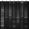
We performed RFLP analysis for six isolated strains of dermatophytes (T. rubrum, T. mentagrophytes var. mentagrophytes, T. mentagrophytes var. interdigitale, T. tonsurans, M. canis, and M. gypseum). From the DNA fragments obtained after BsrDI restriction digestion, we were able to distinguish four different patterns (Table 2). The DNA fragment sizes in each pattern were as follows: T. rubrum, 351/216 base pairs (bp); T. mentagrophytes var. mentagrophytes, 321/216/34 bp; T. mentagrophytes var. interdigitale, 320/216/34 bp; T. tonsurans, 325/216/34 bp; M. canis, 321 bp; and M. gypseum, 428/321/31 bp. Fragments from T. mentagrophytes var. mentagrophytes, T. mentagrophytes var. interdigitale, and T. tonsurans showed an identical pattern because the differences between them were of the order of 1 to 5 bp, which cannot be distinguished by electrophoresis. In addition, T. rubrum, M. canis, and M. gypseum showed their own three distinct patterns (Fig. 2).
DISCUSSION
Dermatophytes are a major cause of frequent fungal infections. In in vitro conditions, morphology of dermatophytes can easily change depending on the culture conditions5. Classification of dermatophytes based on their phenotypes is not systematic. Since the 1980s, molecular classification techniques have been developed to categorize these fungi more accurately6,7,8.
While mitochondrial DNA RFLP analyses (using restriction endonucleases) as well as nuclear ribosomal RNA gene analyses (using nucleic acid sequences) have been attempted9, some species exist that cannot be distinguished using such methods. Since the mid-1990s, analysis of diverse ITS sequences began. Summerbell et al.10 performed the ITS region analysis of T. rubrum and similar strains. They found that T. rubrum, T. raubitschekii, T. fischeri, and T. kanei share similar sequences, and that T. soudanense and T. megninii have nearly identical sequences. Furthermore, several researchers performed an ITS sequence analysis using BsYiI, HinfI, DdeI, and MvaI, and they were able to differentiate 13 strains, including T. rubrum, M. canis, and T. mentagrophytes11,12,13,14.
Dermatophytes are known to have evolved from a single ancestor that co-evolved with mammals for 50 million years. Mitochondrial DNA, which differs from the existing genetic information, is translated to RNA, which is then transcribed into amino acids. Eventually, the reconstructed protein differs from the existing protein, resulting in the development of a new species. Metalloproteinase-1 is an essential factor in the differentiation process of these organisms.
Due to the above process of differentiation, the metalloproteinase-1 sequence in each dermatophyte strain is different. Therefore, it has been possible to classify the dermatophyte strains by molecular techniques using metalloproteinase-1 sequence analysis.
In this study, T. rubrum, the main causative agent of superficial fungal infections, was easily differentiated from T. mentagrophytes var. mentagrophytes, T. mentagrophytes var. interdigitale, T. tonsurans, M. canis, and M. gypseum . Moreover, we were able to differentiate M. canis and M. gypseum from the other four species. On the other hand, we could not differentiate T. mentagrophytes var. mentagrophytes, T. mentagrophytes var. interdigitale, and T. tonsurans. These species could be differentiated by the observation of colony morphology in culture and by microscopy. ITS-1 and ITS-2 sequence analysis using 3 to 4 restriction enzymes for identifying dermatophytes has been reported previously15. To our knowledge, this is the first report to use metalloproteinase-1 sequence analysis for the differentiation of dermatophytes. Various types of proteases such as metalloproteinase-1, metalloproteinase-2, metalloproteinase-9, and metalloproteinase-12 have been previously reported. The use of metalloproteinase DNA sequences for molecular analysis of dermatophytes might lead to improvements in the understanding of their epidemiology and pathogenesis. Furthermore, our current method of categorizing dermatophytes can contribute to more efficient genotyping analysis.

 XML Download