

 PDF
PDF ePub
ePub Citation
 Print
Print


INTRODUCTION
The skin, composed of epidermis, dermis, and subcutaneous tissue, is one of the most active organs in the human body. The epidermis, the outermost barrier tissue, is composed of keratinocytes, melanocytes, and immune cells such as Langerhans cells and γδ T cells1. Epidermal cells actively communicate with each other through a variety of soluble signaling factors (growth factors, cytokines, chemokines, and inflammatory mediators) and through direct contact mediated by cell surface molecules. γδ T cells are the first T cells that emigrate from the thymus, and many of these γδ T cells take residence in epithelial tissues, including the skin, intestine, lung, and reproductive tract2. Epidermis-resident γδ T cells are derived from fetal thymic precursor cells and move to skin before birth3. Evidence from multiple laboratories indicates that epidermal γδ T cells play specialized roles in the maintenance of epithelial homeostasis, wound healing, tumor surveillance, infection, and inflammation1,2,3,4,5,6,7,8. These γδ T cells have been shown to express interleukin (IL)-2, IL-3, granulocyte-macrophage colony stimulating factor (GM-CSF), interferon-γ (IFNγ), tumor necrosis factor-α, chemokine (C-C) ligand (CCL)-3 (macrophage inflammatory protein [MIP]-1α), CCL-4 (MIP-1β), CCL-5 (RANTES), and chemokine (C motif) ligand 12,5,6. Similar to chemokines, these cytokines are detected at low levels under resting conditions, but are rapidly upregulated upon stimulation. The ability of epidermal γδ T cells to produce numerous cytokines and chemokines suggests that these molecules are key players in the immune responses stimulated by the epidermal γδ T cells.
Epidermal γδ T cells are significantly different from those of the peripheral blood in terms of ontogeny, tissue tropism, and antigen receptor diversity6,9. In humans, most of the peripheral blood γδ T cells express the Vδ2 gene segment10. Peripheral γδ T cells recognize, expand, and release cytokines in response to non-peptide antigens, which are mostly of microbial origin. In contrast, most γδ T cells in epithelial tissues express the Vδ1 gene segment and respond to poorly defined self-antigens expressed by stressed cells3,11,12,13,14. In addition, epidermal γδ T cells primarily possess tissue-specific T cell receptors (TCRs) with limited or no diversity compared to the diverse TCRs expressed by γδ T cells found in the peripheral lymphoid organs and blood13. However, it is unclear whether human epidermal γδ T cells exert any functions different from those of peripheral γδ T cells resulting from their differential cytokine production profiles.
In the present study, we compared the cytokine profiles of human epidermal and peripheral blood γδ T cells to investigate the differential activities of epidermal γδ T cells, which affect their neighboring epidermal cells.
MATERIALS AND METHODS
Human skin specimens and blood samples
Skin and peripheral blood samples were collected from human volunteers after informed consent had been obtained according to the approval of the Institutional Review Board at Seoul National University Hospital (IRB No. H-1012-054-344).
Preparation of human epidermal γδ T cells
Whole skin specimens larger than 3×5 cm2 were obtained from three healthy volunteers undergoing elective breast surgery. Skin specimens were incubated in RPMI media containing 2.4 U/ml dispase II (Roche Applied Science, Indianapolis, IN, USA) overnight at 4℃. Epidermal sheets were separated from the dermis by using forceps, and then cultured in complete media (RPMI 1640 media supplemented with 2 mM L-glutamine, 100 U/ml penicillin, 100 mg/ml streptomycin, 10% heat-inactivated fetal calf serum, 100 mM nonessential amino acids, 25 mM 4-(2-Hydroxyethyl)piperazine-1-ethanesulfonic acid (HEPES), 1 mM sodium pyruvate, and 50 mM 2-b-mercaptoethanol in the presence of 50 U/ml IL-2 at 37℃ in a 5% CO2 incubator for 3 to 4 days. Epidermal sheets were then gently agitated to isolate the epidermal cells. Harvested epidermal cell suspensions were enriched for T cells by using Histopaque 1077 (Sigma-Aldrich, St. Louis, MO, USA) gradient centrifugation at 400×g for 30 minutes at room temperature. Epidermal γδ T cells were selected from epidermal mononuclear cell suspensions by using the TCRγδ+ T Cell Isolation Kit (Miltenyi Biotec, Bergisch Gladbach, Germany) and a magnetic cell separator, according to the manufacturer's instructions.
Preparation of human peripheral blood γδ T cells
Human peripheral blood mononuclear cells were isolated from peripheral blood, collected from the antecubital veins of three healthy volunteers, by performing Histopaque 1077 gradient centrifugation at 400×g for 30 minutes at room temperature. Peripheral γδ T cells were selected using the TCRγδ+ T Cell Isolation Kit and a magnetic cell separator, according to the manufacturer's instructions.
Flow cytometry
The purity of γδ T cells was analyzed by flow cytometry using fluorescein isothiocyanate-conjugated anti-human CD3 monoclonal antibodies (mAbs; BD Biosciences, Bergisch Gladbach, Germany), APC-conjugated anti-human TCRγδ complex mAbs (BD Biosciences, San Jose, CA, USA), or the corresponding fluorescently conjugated isotype-matched control Abs. Two-color flow cytometry was performed using the BD FACSCalibur Flow Cytometer, and the results were analyzed using the Cell Quest software (BD Biosciences, USA).
Cytokine array
Epidermal and peripheral blood γδ T cells were stimulated using immobilized 10 mg/ml anti-human CD3 mAb (BD Biosciences, USA) and 100 U/ml IL-2 plus 500 ng/ml phytohaemagglutinin for 36 hours. The supernatants of the cultures were analyzed using the Proteome Profiler Human Cytokine Array Kit, Panel A (R&D systems, Minneapolis, MN, USA), according to the manufacturer's instructions. The cytokine blot intensities on the array membranes were measured using the ChemiDoc gel documentation system (Bio-Rad, Hercules, CA, USA). The relative intensities of the blots were calculated using the following formula: 100×(intensity of sample-intensity of negative control)/(intensity of positive control-intensity of negative control). Data are presented as means±standard deviation.
Enzyme-linked immunosorbent assays
IL-4 and IL-13 levels were measured in the culture supernatants by using the OptEIA™ human IL-4 enzyme-linked immunosorbent assay (ELISA) set and human IL-13 ELISA set (BD Biosciences, USA), respectively, according to the manufacturer's instructions.
Statistical analysis
Data were analyzed using the Student's t-test to determine the significant differences between the cytokine levels in epidermal γδ T cells versus those in peripheral γδ T cells. SPSS ver. 15.0.1. software (SPSS Inc., Chicago, IL, USA) was used for statistical analyses. A p-value of <0.05 was considered statistically significant.
RESULTS
Comparison of the cytokine expression profiles of epidermal and peripheral blood γδ T cells
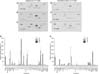
The blot intensities on the cytokine array membranes from activated epidermal (Fig. 2A~C) and activated peripheral blood γδ T cells (Fig. 2D~F) were measured as described in the Materials and Methods. A summary of these data is presented in Fig. 2G (epidermal γδ T cells) and Fig. 2H (peripheral blood γδ T cells). These data showed that both epidermal and peripheral blood γδ T cells produced comparable levels of GM-CSF, I-309 (CCL-1), IFNγ, macrophage migration inhibitory factor, MIP-1α (CCL-3), and RANTES (CCL-5). In addition, we found that epidermal γδ T cells produced significantly higher levels of IL-4, IL-8, IL-13, and MIP-1β than peripheral blood γδ T cells did (p<0.05).
Quantitative analyses of interleukin-4 and interleukin-13 production in epidermal and peripheral blood γδ T cells
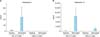
The ELISA results presented in Fig. 3 confirmed that epidermal γδ T cells produced significantly higher levels of IL-4 and IL-13 than peripheral blood γδ T cells did. At the same time, epidermal γδ T cells produced IL-13 at levels several hundred-fold higher than the levels of IL-4 produced.
DISCUSSION
In this study, we analyzed and compared the cytokine expression profiles of human epidermal and peripheral blood γδ T cells to determine the specific activity of epidermal γδ T cells in modulating skin immune responses.
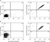
Although activated epidermal γδ T cells are a rich source of several cytokines and chemokines, the transcripts of IL-1α, IL-1β, IL-6, IL-7, IL-10, and IL-13 are in low abundance, and intracellular cytokine staining fails to detect IL-4, IL-5, and IL-1015,16. Thus, it was unexpected that we found that activated human epidermal γδ T cells produced significant levels of both Th1-type cytokines (IFNγ) and Th2-type cytokines (IL-4, IL-5, and IL-13) at the protein level (Fig. 2A). Next, we checked the purity of the cells to confirm that IL-4 and IL-13 were produced by γδ T cells and not by other cells contaminating the cultures. Our flow cytometry results showed that 98.1% and 90.6% of epidermal and peripheral blood cells, respectively, were CD3+TCRγδ+ (Fig. 1).
Interestingly, our results showed that activated epidermal γδ T cells produced significantly higher levels of IL-4, IL-8, IL-13, and MIP-1β than peripheral blood γδ T cells (Fig. 2). IL-8 and MIP-1β are well-known chemotactic factors that are produced from epithelial cells as well as from lymphocytes. In this study, we focused on the predominant cytokines produced by T cells, IL-4 and IL-13, which are typical Th2-type cytokines17. These cytokines affect a variety of cell types including T cells, B cells, natural killer cells, mast cells, monocytes/macrophages, endothelial cells, epithelial cells, dendritic cells, fibroblasts and keratinocytes18,19. In the skin, IL-4 and IL-13 are known to play multiple roles in diseases such as atopic dermatitis (AD) and vitiligo20,21. The concentration of IL-13 is reported to be abnormally high in the serum of AD and vitiligo patients22,23,24,25, and the acute skin lesions of AD patients contain increased numbers of cells expressing IL-4, IL-5, and IL-13 mRNA26,27,28. IL-4 might be important during the initial phase of allergic responses and in the priming and development of Th2 cells, whereas IL-13 plays a more critical role in immunoglobulin E induction and chronic AD pathogenesis29. Our quantitative ELISA data confirmed that epidermal γδ T cells produced markedly higher levels of IL-4 and IL-13 than peripheral blood γδ T cells, and the amount of IL-13 produced by the cells was several hundred-fold higher than the levels of IL-4 produced (Fig. 3). In summary, our results suggest that epidermis-resident γδ T cells have stronger potential to participate in Th2-type responses than γδ T cells existing in the periphery. These data suggest that epidermal γδ T cells play an important role in the pathogenesis of Th2-dominant skin diseases through the active production of IL-13.
 XML Download