

 PDF
PDF ePub
ePub Citation
 Print
Print


INTRODUCTION
The hormonally active vitamin D3 metabolite calcitriol (also known as 1,25-dihydroxyvitamin D3 or 1,25(OH)2D3) has immunomodulatory effects in the skin in addition to its roles in bone metabolism, calcium homeostasis, cell differentiation, proliferation. Calcitriol also cause changes in cytokine expression, suppresses keratinocyte proliferation, promotes keratinocyte differentiation, and induces the expression of antimicrobial peptides (AMPs) such as human β-defensin 3 (HBD-3) and the human cathelicidin family member LL-37.
There is convincing evidence that vitamin D3 directly regulates AMP gene expression in human skin1,2. The promoter regions of the human cathelicidin AMPs (CAMP) and defensin 2 genes contain consensus vitamin D responsive elements (VDREs) that mediate calcitriol-dependent gene expression3, and vitamin D has been shown to enhance the expression of LL-37 in cultured human keratinocytes in vivo1. Induction of CAMP expression in keratinocytes and monocytes is mediated by Toll-like receptors (TLRs)4,5. However, vitamin D3 was shown to down-regulate the expression of TLR2 and TLR4 in a human monocyte in vitro model6, to dose-dependently suppress the protein and mRNA levels of TLR2 and TLR4 in monocytes7. Vitamin D-induced LL-37 up-regulation would therefore be expected to worsen inflammation in psoriasis; however, vitamin D analogs have long been used in the topical treatment of psoriasis. While one study demonstrated that the vitamin D analog calcipotriol suppressed the expression of HBD-2 and LL-37 induced by lipopolysaccharide (LPS) or ultraviolet B (UVB) irradiation in cultured human keratinocytes8, the molecular effects of vitamin D on TLRs and AMP such as LL-37 have not been elucidated in keratinocytes.
For this reason, we sought to determine the effects of calcitriol on the expression of TLR2, TLR4, LL-37 in cultured human keratinocytes. Furthermore, we performed cytokine analysis for tumor necrosis factor α (TNF-α) and interleukin 1β (IL-1β) to confirm the effects of calcitriol on TLR2 and TLR4.
MATERIALS AND METHODS
Cell isolation and culture
For harvesting normal human keratinocytes (NHKs), neonatal foreskin was obtained from neonatal circumcision specimens and primary culture was carried out. Briefly, neonatal foreskin was chopped into 1-mm pieces and trypsinized at room temperature overnight. After vortexing the sample vigorously and incubating for 5 minutes, the supernatant was plated in 25-cm2 culture flasks and incubated in 5% CO2 at 37℃ in keratinocyte growth medium (KGM; Clonetics, East Rutherford, NJ, USA) containing growth supplements and a calcium concentration of 0.03 mM. After 4 passages, cultured keratinocytes were plated at 2×10⁵ cells/ml in a standard flat-bottomed plate. The keratinocytes were starved overnight in keratinocyte basal medium supplemented with serum-free KGM.
The cells were divided into six groups as follows: negative control group, LPS-treated group (5 µg/ml; Sigma, St. Louis, MO, USA), UVB-irradiated group (20 mJ/cm2), calcitriol-treated group (10 nM; Sigma), LPS plus calcitriol-treated group, and UVB plus calcitriol-treated group. The LPS plus calcitriol-treated group was treated with 10 nM calcitriol 30 minutes after the LPS treatment, and the UVB plus calcitriol-treated group was treated with 10 nM calcitriol 30 minutes after the UVB irradiation and incubated for 24 hours.
Ultraviolet B irradiation
The UVB irradiation of 20 mJ/cm2, which was chosen based on preliminary data, was delivered with a Philips TL 20W/12 (Philips, Eindhoven, Netherlands) fluorescent bulb emitting 280 to 320 nm wavelengths with a peak at 313 nm. Before UVB irradiation, the medium was removed and replaced with phosphate-buffered saline. Irradiation output was monitored with a Waldmann UV-meter (Waldmann, Villigen-Schwenningen, Germany).
Preparaton of primers
Polymerase chain reaction (PCR) primers were designed based on Gene Bank (www.ncbi.nlm.nih.gov) data using a DNA synthesizer (Pharmacia; Bjorkgatan, Uppsala, Sweden). The sequences were as follows:
-
TLR-2 (298bp)
5'-GGC CAG CAA ATT ACC TGT GT-3' (sense) and5'-TTC TCC ACC CAG TAG GCA TC-3' (anti-sense); -
TLR-4 (167bp)
5'-TGA GCA GTC GTG CTG GTA TC-3' (sense) and5'-CAG GGC TTT TCT GAG TCG TC-3' (anti-sense); -
TNF-α (219bp)
5'-CAG AGG GCC TGT ACC TCA TCT GA-3' (sense) and5'-GGA AGA CCC CTC CCA GAT AG-3' (anti-sense); -
IL-1β (205bp)
5'-GGG CCT CAA GGA AAA GAA TC-3' (sense) and5'-TTC TGC TTG AGA GGT GCT GA-3' (anti-sense); -
LL-37 (183bp)
5'-GCT AAC CTC TAC CGC CTC CT-3' (sense) and5'-GGT CAC TGT CCC CAT ACA CC-3' (anti-sense); -
GAPDH (238bp)
5'-GAG TCA ACG GAT TTG GTC GT-3' (sense) and5'-TTG ATT TTG GAG GGA TCT CG-3' (anti-sense).
Reverse transcription-polymerase chain reaction
Total RNA was isolated from one dish of cultured keratinocytes using 1 ml of TRIzol reagent (Invitrogen, Carlsbad, CA, USA). After 5 minutes at room temperature, 0.2 ml of chloroform/ml of TRIzol reagent was added, tubes were shaken vigorously by hand for 15 seconds, incubated at 15℃ to 30℃ for 3 minutes. The mixtures were centrifuged at 12,000 rpm (14,000 g) at 4℃ for 15 minutes, the upper aqueous phase was transferred to a fresh tube, and an equal amount of 2-propanol was added. After mixtures were incubated at 4℃ for 15 minutes, they were centrifuged at 12,000 rpm at 4℃ for 15 minutes. The supernatant was removed, and RNA pellets were washed with 500 µl of 70% ethanol, centrifuged at 12,000 rpm at 4℃ for 5 minutes, briefly dried. The purified RNA was dissolved in 30 µl of diethyl pyrocarbonate-distilled water. Three micrograms of total cellular RNA was reverse transcribed at 42℃ for 30 minutes in a 20 µl volume containing 1 µl of reverse transcriptase (TaKaRa; Shiga, Japan), 2 µl of 10× buffer, 2 µl of 10 mM dNTP, 1 µl of oligo dT primer solution, 0.5 µl of RNase inhibitor and 4 µl of 25 mM MgCl2. Two microliters of each resulting cDNA sample was amplified by PCR in 25 µl containing 2.5 µl of 10× buffer, 2.5 µl of 25 mM MgCl2 and 0.75 µl of 10 pmol primer solution.
Thermal cycle profiles were conducted using the following conditions: 94℃ for 5 minutes, 35 cycles of 94℃ for 1 minute, 59℃ for 1 minute, 72℃ for 1 minute and a final extension step of 72℃ for 10 minutes.
Electrophoresis
The PCR products were run on a 1.5% agarose gel, separated by electrophoresis for 15 minutes at 100 volts, and visualized by UV transillumination.
Real-time polymerase chain reaction
RNA was isolated using TRIzol Reagent (Invitrogen), and cDNA was synthesized using the RevertAid™ First Strand cDNA Synthesis Kit (Fermentas, California, MD, USA). The primer sequences for GAPDH, TLR2, TLR4, TNF-α, IL-1β and LL-37 were the same as those described above. The purity and quantity of each sample were determined by UV absorption and gel electrophoresis. Real-time PCR of target cDNA was conducted for TLR2, TLR4, TNF-α, IL-1β and LL-37, and normalized to GAPDH gene expression. All SYBR Green reactions used SsoFast™ Eva-Green (BioRad, Mississauga, ON, Canada). Real-time PCR amplification was performed on a CFX96™ Real-Time System (BioRad).
Enzyme-linked immunosorbent assay
Cell culture supernatants were collected after drug treatment, centrifuged to remove cellular components, and stored at -80℃. To determine the amount of TNF-α in each supernatant, antibodies directed against human TNF-α were used as the capture and detection antibodies. The fluorescent substrate horseradish peroxidase-avidin (R&D Systems, Baltimore, MD, USA) was used for color development. The amount of cytokine in the test sample was determined from standard curves established with serial dilutions of recombinant human TNF-α (R&D Systems). TNF-α concentrations were measured with the SpectraMax 340PC384 System (Molecular Devices, Wokingham, UK).
Statistical analysis
Data are presented as mean±standard deviation. Multiple comparisons were adjusted according to ANOVA. p-values are two-sided and p<0.05 was considered statistically significant. All statical analyses were performed by using a Sigma Plot for Windows (Systat software Inc., San Jose, CA, USA).
RESULTS
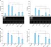
Increased TLR2 and TLR4 mRNA expression is suppressed by calcitriol
Calcitriol may act through various pattern recognition receptors9. Hence, we investigated whether various pattern recognition receptors such as TLR2 and TLR4 could be influenced by calcitriol in NHKs. Because there was no change in the expression of TLR2 and TLR4 when NHKs were treated with calcitriol (10 nM), the keratinocytes were stimulated with LPS (5 µg/ml) or UVB irradiation (20 mJ/cm2). As expected, mRNA levels of TLR2 and TLR4 were found to be increased as much as 30 times (Fig. 1). This effect was suppressed when the cells were treated with calcitriol (10 nM) prior to stimulation with LPS or UVB irradiation (20 mJ/cm2) (Fig. 1). These differences were statistically significant relative to the control group (p<0.05).
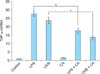
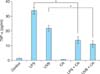
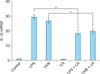
Calcitriol suppresses over-expressed proinflammatory cytokines
We investigated the effects of calcitriol on the immune response by stimulating keratinocytes in the presence of 10 nM calcitriol and measuring the cytokine levels in the cells and supernatant. The expression of TNF-α mRNA in NHKs was up-regulated 24 hours after stimulation with LPS or UVB irradiation. This effect was diminished upon treatment with calcitriol (Fig. 2). The levels of supernatant TNF-α were also up-regulated upon stimulation with LPS or UVB irradiation. However, the amount of secreted TNF-α was reduced after calcitriol treatment (Fig. 3). The expression of IL-1β mRNA in NHKs was also up-regulated upon stimulation with LPS or UVB irradiation, and this effect was diminished after treatment with calcitriol (Fig. 4).
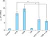
Increased LL-37 mRNA expression is suppressed by calcitriol
LL-37 mRNA expression was not detected in the cultured keratinocytes of the unstimulated controls. LL-37 mRNA expression was increased in the UVB-irradiated and LPS stimulated groups, and decreased after treatment with calcitriol (Fig. 5). These effects were statistically significant when compared with the results of the control group (p<0.05).
DISCUSSION
In this study, we investigated the effect of the vitamin D analog calcitriol on the expressions levels of TLR2, TLR4, and LL-37 in cultured human keratinocytes. The mRNA levels TLR2 and TLR4 were up-regulated in keratinocytes stimulated with LPS or UVB irradiation, while this effect was diminished after treatment with calcitriol. TLRs play important roles in the innate immune response to microbial infection. Dysregulation of TLR signaling is linked to a number of disease conditions, and possible roles for TLRs in innate immunity activation in psoriasis have been investigated. Recent studies have suggested that TLR2, TLR4, and γ δ T-cell receptors may recognize heat shock protein 60 as a ligand and consequently activate the immune system10,11,12.
TLRs signal via the transcription factor nuclear factor-κB, which regulates the transcription of proinflammatory cytokines such as TNF-α, IL-1 and IL-6. For this reason, we performed cytokine analysis for TNF-α and IL-1β to confirm the effects of calcitriol on TLRs. The expression levels of TNF-α and IL-1β in keratinocytes were up-regulated upon stimulation with LPS and UVB irradiation. This effect was diminished after treatment with calcitriol.
Secretion of TNF-α was also suppressed after treatment with calcitriol. TNF-α is a pluripotent but predominantly proinflammatory cytokine, and is a major factor in the early steps of the innate immune response. TNF-α production is induced by LPS/lipoteichoic acie stimulation through TLR2- and TLR4-dependent pathways. Therefore, TNF-α production might be inhibited upon TLR inhibition. Calcitriol would be expected to suppress TNF-α synthesis through the down-regulation of TLRs in keratinocytes. Our results imply that calcitriol is able to modify the cytokine response towards an anti-inflammatory profile. This effect of calcitriol on the expression in TNF-α in keratinocytes could be one mechanism of its of action mechanisms in the treatment of psoriasis.
The link between TLR activation and the expression of AMPs is clearly established5. Kumar et al.13 demonstrated that expression of HBD2 is regulated by TLR2-dependent pathways. Recently, the immunomodulatory effect of calcitriol through down-regulation of TLR2 and TLR4 expression was demonstrated in a human monocyte in vitro model6. Do et al.7 showed that vitamin D3 was found to dose-dependently suppress the protein and mRNA levels of TLR2 and TLR4. Based on these results, calcitriol would be expected to suppress TLRs, and in turn, TLR-mediated AMP expression in keratinocytes. The present study also showed that LL-37 induction was suppressed after treatment with calcitriol. Although we did not confirm the pathway by which calcitriol affected AMP expression, our results suggest a possible mechanism in which calcitriol suppresses TLR activation and in turn decreases TLR-mediated AMP expression in keratinocytes. These results suggest that calcitriol may exert its therapeutic effect on psoriasis by regulating TLR2- and TLR4-mediated AMP expression in keratinocytes.
Several recent reports suggest a connection between vitamin D3 and AMP expression in keratinocytes. In the presence of vitamin D, human keratinocytes upregulate LL-37 expression in response to TLR2 and IL-17 signaling4,14. Vitamin D analogs activate vitamin D receptor, which in turn would be expected to bind the VDREs in the promoter regions of cathelicidin genes and thus increase LL-37 expression3. Increased LL-37 would then aggravate inflammation by binding self-DNA and activating plasmacytoid dendritic cells in psoriasis15,16. However, in reality, opposite is true: indeed, vitamin D analogs are a mainstay in the topical treatment of psoriasis. To date, mechanisms that could explain this paradoxical effect of vitamin D analogs on psoriasis are not completely understood. Recent publications highlight the role of dysregulated AMP expression in the pathogenesis of psoriasis. HBD and LL-37 levels are greatly increased in the keratinocytes of psoriatic plaques17. It was demonstrated that LL-37 is able to suppress the induction of apoptosis in keratinocytes18. Therapeutic approaches to restore normal LL-37 expression in keratinocytes may prove beneficial for the treatment of psoriasis. Our previous study8 demonstrated that calcipotriol decreased the expression of HBD-2 and LL-37 induced by UVB and LPS in cultured human keratinocytes. The present study demonstrated that calcitriol suppress the induction of LL-37 and proinflammatory cytokines in addition to TLR2 and TLR4 upon LPS and UVB radiation treatment of cultured human keratinocytes.
In conclusion, calcitriol was found to suppress the LPS- and UVB-mediated induction of TLR2 and TLR4 in human keratinocytes. This study suggests that calcitriol modulates the expression of AMPs or TNF-α in chronic inflammatory skin diseases associated with overexpression of these factors. Although our experimental results are in conflict with the current understanding of vitamin D effects on AMPs and proinflammatory cytokines, our results support the application of calcitriol in therapy for psoriasis and the possibility for alternative pathways of vitamin D analogs activity in psoriasis patients.
 XML Download