

 PDF
PDF ePub
ePub Citation
 Print
Print


INTRODUCTION
Large-area skin defects and deep burns are difficult to treat with satisfactory clinical results. Although a variety of skin substitutes currently used in the clinic have some effects in the early period of treatment of severe scald or burn injuries, they cannot be used as permanent skin substitutes owing to their lack of dermal and epidermal structures and skin appendages1,2,3. Stem cell technology along with the development of tissue engineering technology can now provide new ways to promote wound healing. Many researchers are beginning to develop tissue-engineered skin for repairing skin defects and scald wounds4,5,6. The key to tissue engineering is the choice of a scaffold and seed cells. Bone marrow mesenchymal stem cells (BMSCs) can differentiate into a variety of tissues, including tendons and ligament, as well as cells, including osteoblasts, cartilaginous cells, and nerve cells7. Owing to the ease of isolation, culture, and proliferation, as well as the low rate of graft rejection of BMSCs, these cells are now the focus of cell biology and tissue engineering research. Fibrin glue has been used widely in biomedical fields as a natural macromolecular material with excellent biocompatibility8,9,10,11. In the present experiment, we chose rat BMSCs as the seed cells and fibrin glue as the scaffold. Fibrin glue and rat BMSCs were mixed to produce an active dressing film, which was used to repair burn wounds on the skin of rats. We expected the mixture to promote skin tissue repair and the generation of skin appendages.
MATERIALS AND METHODS
Animals
All experiments were conducted in conformity with the institutional guidelines of the Zhejiang Academy of Medical Sciences, Hangzhou, China, and the National Institutes of Health Guide for Care and Use of Laboratory Animals for the care and use of laboratory animals. Four 1-day-old Sprague-Dawley (SD) rats were used as the sources of BMSCs, and 15 adult rats were used to make scald animal models for allogeneic BMSC transplantation. The mean weight of the adult rats was 150 g. For histological analyses, 15 rats were killed 30 days after transplantation.
Cell isolation and culture
Isolation of primary rat BMSCs was performed as follows: 1-day-old SD rats were anesthetized and decapitated to obtain the femur and tibia with a sterile operation. After being washed with phosphate buffered saline (PBS) containing 1% (v/v) penicillin and gentamicin, the tissues were chopped into small pieces and immersed in 2 ml of low-glucose Dulbecco's modified Eagle's medium (LG-DMEM, Invitrogen; Gibco, Grand Island, NY, USA). After complete mixing, the obtained suspension was filtered through a 200-mesh sieve. The cell suspension was plated on 24-well culture plates (Corning, Corning, NY, USA) with complete culture medium (LG-DMEM supplemented with 10% [v/v] fetal bovine serum [FBS], 100 U/ml of penicillin, and 100 mg/ml of gentamicin), which was incubated at 37℃ with 5% of humidified CO2. The culture medium was changed every 2 days. Cells within the sixth passage were used in the present study.
In vitro differentiation assay
For chondrocyte differentiation, fourth-passage cells were cultureded on a 3.5-cm-diameter dish (Corning) with complete culture medium of LG-DMEM until the cells reached 80% confluence. Then, the medium was replaced with a chondrogenesis medium comprising high-glucose DMEM (HG-DMEM, Invitrogen) supplemented with 10 µg/L of transforming growth factor-β1 (PeproTech, Rocky Hill, NJ, USA), 4×10⁻⁵ g/L dexamethasone (Sigma-Aldrich, St. Louis, MO, USA), 0.05 g/L of ascorbic acid, 4.32 g/L of β-glycerophosphate, 2×10⁻³ g/L of insulin (Sigma-Aldrich), 0.10 g/L of pyruvate, 1.50 g/L of bovine serum albumin (BSA), 3% (v/v) of FBS, 1% (v/v) of penicillin and gentamicin. The medium was changed every 2 days. After 28 days of culture, the BMSCs were stained with toluidine blue O (Sigma-Aldrich) and observed by using microscopy.
For osteoblast differentiation, the cells were cultured in HG-DMEM supplemented with 4×10⁻⁵ g/L of dexamethasone, 0.05 g/L of ascorbic acid, 4.32 g/L of β-glycerophosphate, 3% (v/v) of FBS, and 1% (v/v) of penicillin and gentamicin. The BMSCs were cultured in osteogenic medium for 21 days and then stained with von Kossa dye to detect the deposition of mineralized nodules.
Determination of cell surface antigens
A 100-µl suspension of BMSCs with a density of 1×10⁶ cells/ml was added to tubes for flow cytometry analysis (FACSCalibur; BD Biosciences, Franklin Lakes, NJ, USA). A total of 2 µl of anti-rat CD29-FITC (cluster of differentiation 29-fluorescein isothiocyanate), 4.5 µl of anti-rat CD106-PE (CD106- phycoerythrin), 0.5 µl of anti-rat CD44H-FITC, and 0.5 µl of anti-rat CD90/mouse CD90.1-FITC (Biolegend, San Diego, CA, USA) were each added to the tubes. After incubating the mixture with the antibodies for 20 minutes at room temperature and storing away from light, the mixtures were rinsed with 1 ml PBS in the presence of 1% BSA and resuspended into 500 µl PBS to determine the cell surface antigens. The results were analyzed by using CellQuest software (BD Biosciences) and the unlabeled rat BMSCs were used as the negative control.
Preparation of the rat model with scalded skin and the experimental groups
A total of 15 SD rats with a mean weight of 150 g were enrolled in this study. The rats were intraperitoneally anesthetized with 3% (m/v) pentobarbital sodium. The back hairs of the rats were clipped. Then, the rats were placed in a prone position on the operation table and their extremities were tied. The back of the animals was scalded for 20 seconds by using a beaker containing half a cup of boiling water. Two scald wounds were made on the backs of the rats, one on each side. After scalding, each rat was treated with an intraperitoneal injection of 2 ml of physiological saline.
A total of 30 scald wounds on the rats' backs were randomly divided into 3 groups with 10 scald wounds each: (1) experimental treatment group; (2) experimental control group; and (3) blank control group.
Transplantation of the marrow mesenchymal stem cell/fibrin glue complex into scald wounds
The rats were subjected to escharotomy on the fourth day after scalding, and different treatments were conducted for the 3 groups. In the experimental treatment group, the scald wounds were covered with the mixture of fibrin glue and BMSCs by using a 'Y'-type syringe. The gel that formed within 2 minutes contained approximately 5×10⁷ transplanted cells. The wounds of the experimental control group were injected only with fibrin glue. No intervention was administered to the blank control group. After transplantation, all wounds were bandaged for 3 days and then the bandages were removed. Observation and measurement of the diameters of the scald wounds were performed 30 days after treatment. The scald local tissues of all rats from the 3 groups were cut into pathological sections, stained with hematoxylin and eosin, and observed under the microscope (Olympus Corporation, Tokyo, Japan).
Statistical analyses
The experimental data were reported as mean±standard deviation; the differences of the diameters of the scald wounds in the 3 groups were analyzed statistically by using the t-test. All statistical analyses were performed using IBM SPSS Statistics 20.0 (IBM Co., Armonk, NY, USA) and p-values of <0.05 were considered significant.
RESULTS
Morphology and proliferation activities of marrow mesenchymal stem cells
On the first day of culture, the attached cells were few in number and distributed as short spindle-like cells. On the next day, many microcolonies appeared and the cells became elongated. The colonies multiplied quickly and gradually converged together. The cells showed a uniform, long spindle shape, and they were arranged spirally. On the fifth day, a cell monolayer was observed. The subcultured cells were initially spherical, and quickly attached to the wall of the plates within several hours and assumed a spindle shape again. After 3 days of cell transfer, a cell monolayer was observed. No significant changes were observed after 5 serial passages; however, increasing the number of passages would lead to diverse cell forms and aging cells.
The sixth-generation BMSCs were found to be normal diploid cells (1.00), with 86.99% of the cells in the G0/G1 phase. This showed that the cells had good proliferation activity (Fig. 1A).
Identification of marrow mesenchymal stem cells
After 21 days of osteogenic induction, the cells were positively stained with the von Kossa dye. Deposition of many brown focal mineralized nodules was observed. BMSCs that were chondrogenically induced for 28 days stained positive with toluidine blue dye, whereas the control groups showed negative staining. These results suggest that the BMSCs were pluripotent (Fig. 1B).
The expression of CD29, CD44H, CD90, and CD106 as surface markers of BMSCs was detected by using flow cytometry. All markers were positive, with the rate of positive expression being 99.58%, 93.30%, 82.50%, and 45.43% for CD29, CD44H, CD90, and CD106, respectively. The results were compatible with the characteristics of mesenchymal stem cells.
Rat scald model
1) General observations
The scalded skin area was pale, and the skin became raised 10 minutes after scalding. On the first day after scalding, the skin gradually became red and some exudation appeared. On the fourth day after scalding, an eschar appeared on the scalded area, as well as yellow exudation around the scald. Physical activity of the rats significantly reduced (Fig. 2).
2) Pathological observations
The pathological sections of the eschar showed that the epithelial cells had nuclear pyknosis. The collagen and elastic fibers in the dermal layer had been destroyed. A few residual hair follicles were visible, and the sebaceous glands showed structural failure. These histopathological results were compatible with skin pathological changes in deep second-degree burns12, suggesting that the method of making the rat scald model was feasible.
Transplantation therapy for the scalded rats
1) General observations
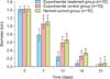
On the third day after transplantation, the wound dressings were removed. No substantial wound contraction was observed in all 3 groups. The mean diameter of the wounds was approximately 1.42 cm, and the wounds appeared bright red. On the seventh day after transplantation, the mean diameter of the dry and dark red wounds considerably reduced to approximately 0.79 cm in the experimental treatment group. However, the mean diameter of the wounds in the experimental control group and the blank control group was approximately 1.07 cm and 1.12 cm, respectively. The wounds of the experimental control group appeared dry and dark red, and a gray exudate remained on the wounds of the blank control group. On the 12th day after the transplantation, the mean diameter of the wounds was 0.25 cm in the experimental treatment group, and approximately 0.59 cm and 0.67 cm for the experimental control group and the blank control group, respectively. On the 14th day after the transplantation, some wounds in the experimental treatment group had healed, whereas the other 2 groups did not show any wound healing. On the 21st day after the transplantation, all the wounds of the experimental treatment group showed healing except for 1. The other 2 groups did not show healing of all the wounds until 30 days later (Fig. 3).
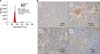
2) Pathological observations
The histopathological sections showed that the newly formed epidermis with a normal structure was thicker than the normal epidermis in all groups. The newly formed dermal layer showed only collagen fibers consisting of fibroblasts. We found that the sebaceous glands showed obviously proliferous at the edge of the new tissue and gradually extended to the deep dermal layer of the new tissue in the experimental treatment group. The epidermal layer recessed and formed a hair follicle-like structure in the experimental treatment group but not in the other 2 groups (Fig. 4).
DISCUSSION
Conventional dressings can only be used as a temporary adjuvant therapy for skin burns but not for the treatment of large-area deep burns. The development of skin tissue engineering could offer patients a better quality of tissue repair. Ideally, patients would prefer tissue-engineered skin that not only can cover the wound immediately but also has the function of normal skin tissue.
The skin is mainly composed of the epidermis and dermis, and each layer consists of a variety of cells. Therefore, 1 of the most important aspects of tissue engineering is to choose a multipotent cell as the seed cell that can develop into all cell types of the skin. In this study, we selected BMSCs as seed cells because they could differentiate into various cells and tissues7,13. Many studies have found that BMSCs can differentiate into cells of the skin appendages14,15, such as sweat gland tissue16, at the wound site. BMSCs can also be induced to express the endothelial cell phenotype under suitable conditions17,18,19. Our experimental results also confirmed that these cells have multiple differentiation potentials. BMSCs have low immunogenicity. They can secrete certain cytokines to activate CD4+/CD25+ regulatory T-cells, inducing immune tolerance20,21,22. Therefore, in our experiments, we used allogeneic cells for transplantation to solve the problem of insufficient cells.
Another important aspect is the choice of scaffolds for the tissue-engineered skin. Fibrin glue is extracted from human plasma. The biological product is mainly composed of fibrinogen and thrombin preparations. It imitates the final stage of blood coagulation, that is, with the action of thrombin, Ca2+, and clotting factor XIII, fibrinogen rapidly solidifies and forms fibrin glue as a high-porosity structure consisting of a 3-dimensional network. The pore structure and high porosity of fibrin glue make it difficult to wash away by body fluids, and not easily degradable. The structure is also beneficial because it has a variety of growth factors and cells to adhere to23. Fibrin glue also has the characteristic of high plasticity24. It can be made into any shape for use in complex clinical cases, especially for the repair of uneven wounds. Furthermore, the mechanical properties of fibrin glue are superior to those of other gel-like scaffolds. In 2006, Liu et al.25 first reported in 'Science' that fibrin glue is 1 of the known natural fibers with the highest ductility and elasticity. Because fibrin glue has good cell and tissue compatibility, biodegradability, and no rejection and toxicity, it is a good candidate for tissue engineering of scaffolds.
In this study, we mixed fibrin glue with allogeneic BMSCs to produce an active dressing that can be used to coat burn wounds. The experimental results showed that the active dressing quickly formed a film-like gel on the wound. This method can cover wounds immediately, and thus provides a very good choice for patients with burns who need emergency skin substitutes.
From the transplantation therapy for the scalded rats, we found that the composite could promote healing of the scald wounds. There has been no consensus about the exact mechanism of action of MSCs in wound healing. One hypothesis involves the differentiation of MSCs into neo-tissues. Another hypothesis is that MSCs exert paracrine effects on the wound. Many researchers have suggested that the paracrine process may also be involved in tissue repair26. Our previous experiments showed that the transplanted cells are present in the neo-tissue27. Many studies have also demonstrated that BMSCs have the capacity to differentiate into certain cells in the neo-tissue28,29. This suggests that the transdifferentiation of BMSCs is involved in tissue repair.
Histological examination showed that the skin tissue of rats has a strong self-repair capability. In the case of full-thickness skin defects, it can repair itself to form new skin. The regenerated skin has a complete epidermal structure, although its dermal structure is incomplete. Owing to the lack of elastic fibers, the newly formed skin lacks elasticity and has poor mechanical properties compared with normal skin.
Importantly, the sebaceous glands showed obviously proliferous at the edge of the new tissue in the experimental treatment group. Furthermore, there was a hair follicle-like structure in the epidermal layer. The results suggest that BMSCs can promote skin appendage regeneration, which makes it possible to achieve a compound skin with complete physiological function.
In the comprehensive analysis of the experimental results, we found that the composite of fibrin glue and BMSCs could induce the formation of a complete skin structure and accelerate the healing rate of burned skin. The active dressing film made with fibrin glue and BMSCs is a good choice as a skin substitute for the treatment of patients with burns in the emergency setting.


 XML Download