

 PDF
PDF ePub
ePub Citation
 Print
Print


INTRODUCTION
Keratoacanthoma (KA) is a rapidly growing cutaneous neoplasm deriving from the hair follicle. KA usually occurs on sun-exposed sites in elderly people and has a close clinical and histopathological resemblance to squamous cell carcinoma (SCC)1. KA occurs less often among Koreans than Caucasians1,2. KA exhibits a tendency of spontaneous involution, but it is difficult to predict a tumor progression or an involution. KA can cause local tissue destruction if it is allowed to grow, so an early treatment is recommended. Various treatment modalities have been reported including the radiation therapy, a systemic oral retinoids, and an intralesional application of 5-fluorouracil (5-FU), methotrexate (MTX), or interferon α-2a. However, a surgical excision remains the treatment of choice for the majority of cases of KA.
Surgical excision can result in functional and cosmetic defects when a large or strategically located lesions are treated. An effective nonsurgical treatment would be desirable in such cases. Several case reports have described the successful treatment of KA by an intralesional MTX in Korean patients3,4,5. Patients we treated at our institution experienced a relatively slow treatment response to the MTX injection therapy. No study has systematically examined the response to the MTX treatment in Korean patients. Thus, we reviewed the cases of Korean KA patients treated with the intralesional MTX and analyzed the therapeutic regimens by comparing these patients with the Caucasian patients.
MATERIALS AND METHODS
Patients
The patient data from the Department of Dermatology, Korea University Ansan Hospital from 2004 to 2010 were retrospectively reviewed. The Institutional Review Board of Korea University Ansan Hospital granted the approval for the review of the medical records. We identified seven patients with KA whose treatment involved the intralesional injection of MTX. The present study includes two cases that have been previously reported6. In addition to our data, we included the cases of Korean patients with KA treated with the intralesional MTX found in a KoreaMed, KMbase, and PubMed literature search. In total, we reviewed seven cases from our institution and four additional published cases identified in the available literature. Variables including the patient's age, sex, tumor size, and the location, duration, and the histologic confirmation of the diagnosis, the cumulative MTX dose, the total number of injections, the interval between the injections, the treatment outcome, adverse events, and the total patient follow-up time were identified.
Treatment
We used 0.1 to 1.8 ml of MTX (JW Pharmaceutical, Seoul, Korea) at a concentration of either 12.5 or 25 mg/ml injected at the base of the KA tumor using a 24-gauge needle at each treatment session. Shortly after the MTX injection, a uniform tumor blanching was achieved. We adjusted the injection site, the interval between the injections, and the concentration and the amount of MTX depending on the clinical response. We repeated the injections with an interval that ranged from 4 days to 28 days (average 10 days). When the total tumor necrosis was achieved, the treatment was discontinued.
Assessment
At every visit, two independent dermatologists made a clinical assessment of the tumor resolution by using photographs of the lesions. After the resolution, we continued to follow the patients for tumor recurrence and any adverse effects such as nausea, oral ulcer, dyspnea, or alopecia.
Therapeutic analysis
In order to compare the response to the MTX treatment between Koreans and Caucasians, we analyzed nine cases of KA in Korean patients and nine cases of KA in Caucasian patients. The Korean patients with an insufficient amount of treatment information (n=1) and those with an incomplete response (n=1) were excluded from the analysis. We used the data from a study by Melton et al.7 that reported the treatment response, the total amount of MTX used, the injection interval, and the elapsed time to the tumor regression for Caucasian patients. Other data from the Western sources that did not identify the ethnicity of the patients were excluded from the analysis. To calculate the amount of the injected MTX per unit volume and to adjust for the tumor size, we assumed a spherical lesion and divided the total amount of MTX used by the lesion volume. Due to the small sample size, we applied the Mann-Whitney U-test as a non-parametric test to analyze the data. IBM SPSS Statistics 20.0 (IBM Co., Armonk, NY, USA) program was used for statistical analysis while a value of p <0.05 was considered statistically significant.
RESULTS
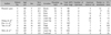
Eleven cases of KA treated with an intralesional MTX in Korean patients were identified: seven cases from our institution and four cases from the published literature (Table 1). At out institution, a pre-treatment biopsy was performed, which confirmed the diagnosis in six patients. The average tumor diameter was 1.7 cm and all tumors were located on the face, a sun-exposed and cosmetically important area.
Among the pooled data of 11 patients, three were male and eight were female. The average age was 62.3 years and the tumors ranged in diameter from 0.5 to 4.0 cm (mean 2.1 cm). A pre-treatment biopsy was done in 82% (9/11) of cases. Most of the tumors (10 of 11, 91%) were located on the face, with one (9.1%) located on the leg. The average total dose of MTX was 53.7 mg per treatment course. An MTX concentration of 25 mg/ml was most common. To achieve the tumor resolution, 2 to 7 injections (mean 4.3 injections) were required. The average injection interval was 10 days (range, 4~28 days). The rate of a complete resolution with an intralesional MTX was 91% (10/11) (Table 1, Fig. 1). In one case, the tumor size decreased after 4 injections of MTX. However, an excisional biopsy was also used to confirm the treatment outcome and exclude the possibility of SCC, which resulted in a complete resection of residual KA lesion.
No significant adverse events occurred in the 11 cases examined. The clinical follow-up ranged from 2 to 84 months, with an average of 18.3 months. No recurrences were noted for the 10 patients for whom the follow up data was available. Scarring occurred rarely and the cosmetic results were good in all patients.
At our institution, seven of the patients received MTX at a concentration of 12.5 or 25 mg/ml at an interval of 4 days to 28 days. Among the four cases from the literature, two cases received MTX at a concentration of 5 mg/ml at an interval of 7 days4. The other two cases received MTX at a concentration of 25 mg/ml at an interval of 7 or 14 days each3,5. In cases where an MTX concentration of 5 mg/ml was used, the injected amount of MTX per tumor size was smaller, the number of the injection was not greater, and the time to the tumor regression was not greater than in cases were an MTX concentration of 12.5 or 25 mg/ml was used. In cases with a relatively short injection interval of 4 days, the time to the tumor regression was not shorter than in cases where the interval was more than 7 or 14 days. Compared with the Caucasian patients in the study of Melton et al.7, in which an MTX concentration of 12.5 or 25 mg/ml at an interval of 14 days was used, the size of KA and the total amount of injected MTX per tumor volume (Korean, 26.7±13.8 mg/cm3 versus Caucasian, 11.0±9.4 mg/cm3) was not significantly different (p >0.05). The cases from the Korean literature showed relatively shorter injection intervals. The Korean patients required 2 to 7 injections (mean 4.6 injections) to achieve a tumor resolution while the mean time to clearing was 7.6 weeks. The Caucasian patients required 1 to 2 injections (mean 1.7 injections) with the mean time to clearing at 3.0 weeks. Compared to the Korean patients, the Caucasian patients showed a tendency to require a less number of injections, and a shorter time to the tumor regression (Table 2).
DISCUSSION
Because KA often regresses spontaneously, some authorities claim that KA does not require a treatment8. However KA may take a long time to involute spontaneously and may continue to enlarge during that time, resulting in an impingement of important structures. In addition, SCC misdiagnosed as a KA may metastasize7. Thus, an early treatment can hasten the cure, prevent functional loss, and improve overall cosmesis9.
Many treatment options are available for KA. Excisional surgery is recommended for the majority of KA cases. However, an excision can cause significant cosmetic or functional problems, because of the surgical defects resulting from the size or location of the tumor7. In one study, 74% (28/38) of KA lesions were located on the face and the scalp10. In our patients, all tumors were located on the face with some tumors exceeding 2 cm in size. In addition, older patients or those with comorbid conditions are not good candidates for a surgical management4. In these cases, an intralesional MTX, as an example of a nonsurgical treatment modality, offers a less invasive treatment option with acceptable cosmetic results (Fig. 1).
MTX is appropriate for rapidly growing tumors since it inhibits DNA synthesis in actively dividing cells. MTX is a folic acid analog that binds to the dihydrofolate reductase, blocking the formation of tetrahydrofolate and preventing the synthesis of the purine nucleotide thymidine11.
In our analysis of the pooled data, the patients sought a medical treatment at the stage of a rapid tumor growth. The time at the start of the treatment ranged from 1 to 3 months from the onset of the disease. Thus, we were unable to examine the efficacy of the intralesional MTX in other stages of KA tumor growth. Intralesional chemotherapy may not be an effective option for slow growing lesions; for example, KA of only 16 weeks duration showed no response to the treatment with an intralesional 5-FU in one study12. Considering the mechanism of action, if an intralesional chemotherapy of non-proliferating KA shows no response, other treatment options are desirable.
In our pooled data from the Korean patients, more injections and a greater amount of MTX were required for the treatment of KA, compared to the previous reports of the patients of other ethnic origins identified in our search of the literature. Eleven Korean patients with an average tumor diameter of 2.1 cm were treated with an average of 4.3 injections and an average total MTX dose of 53.7 mg3,4,5. Melton et al.7 reported that 9 patients, with an average tumor diameter of 1.8 cm, required an average of 1.7 injections and an average cumulative total MTX dose of 21.9 mg. Large KA tumors (>2 cm), which are termed giant KA6, were noted in six of our 11 cases. Nevertheless, the size of KA lesion and the total adjusted amount of applied MTX per unit volume were not significantly different in our patients compared to those from the report of Melton et al.7. The dose, the concentration, and the frequency of the injections were dependent upon the institutional treatment protocol13. We may attribute this to the small number of the cases and the non-standardized treatment protocol.
In the case where an MTX concentration of 5 mg/ml was used, the amount of MTX injected per tumor size was smaller, and the time to the tumor regression was not longer4. In the cases where a shorter interval of treatment was applied, it did not show a faster regression of tumor than those cases where an interval of more than 7 or 14 days was used (Table 1). Thus, one advantage of using MTX is the option to use an injection interval longer than 14 days7,10.
It takes a significantly longer time for the tumor regression in Koreans compared to Caucasians. Although Melton et al.7 insisted that a surgery is necessary if the lesion does not show a complete resolution after two injections of MTX, KA may require a different standard of care. If the tumor responds with a decrease in size, a continuation of injection therapy may be considered regardless of the number of the injection applied.
A further study is required to ascertain whether the observation or the continued regular injections should be considered after the treatment responses such as ulceration or necrosis; it may also help optimize the treatment guidelines for the use of an intralesional MTX for KA based on the size or the location of the tumor.
The complete resolution rate found in this study is consistent with the previous studies using an intralesional MTX for KA. Annest et al.10 reported a cure rate of 92% from a retrospective analysis of 38 patients with KA treated with an intralesional MTX from 1991 to 2006. In our pooled analysis, 91% (10/11) of KA cases showed a complete resolution following the treatment solely with an intralesional MTX; one case showed a marked size reduction and a completely excised residual KA was later confirmed by a post-treatment biopsy. Even if some residual tumor exists after the intralesional injections of MTX, it is still helpful in the surgical management of KA7.
Intralesional injections enable the local delivery of potent chemotherapeutic agents like MTX and can help avoid a systemic toxicity14. In our analysis, none of the patients treated with an intralesional MTX experienced clinically significant adverse events. Two cases of pancytopenia have been reported in intralesional MTX treated patients with a renal insufficiency15,16. Thus, a baseline and a follow-up laboratory monitoring are recommended for patients treated with an intralesional MTX10. We excluded the patients with a medical history of a renal disease.
This small retrospective study has its limitations. First, a residual tumor might remain or the tumor might potentially recur due to the short duration of the follow-up, even though our average follow-up interval was 14 months. In addition, a post-treatment biopsy was not performed. Second, some pathology reports were unable to exclude SCC; it is difficult to differentiate KA and SCC histologically. Thus, we based our diagnosis of KA on the characteristic morphologic features and the rapid pattern of growth6; the response to intralesional MTX was also helpful in confirming the diagnosis.
KA is a rapidly growing tumor that can cause a local tissue destruction. Thus, an early therapeutic intervention is required. Although surgical excision is the treatment of choice, non-excisional treatment is preferred if poor cosmesis and a loss of function is expected from the surgery. Our results, combined with the data from the literature, indicate a nearly 91% complete response rate for an intralesional MTX treatment of KA in Korean patients. This simple procedure is both efficacious and has no significant systemic adverse events. We believe this study could be the first step to developing an efficient treatment algorithm for KA based on the tumor size or the location.


 XML Download