

 PDF
PDF ePub
ePub Citation
 Print
Print


INTRODUCTION
Extramammary Paget's disease (EMPD) is an uncommon intraepithelial adenocarcinoma, which primarily affects the apocrine-bearing skin. EMPD resembles mammary Paget's disease clinically, as well as histopathologically. Bowen disease is equal to that of squamous cell carcinoma in situ, having the potential to progress to squamous cell carcinoma. Its histopathologic features include full-thickness epidermal atypia with adnexal involvement. We experienced a case of EMPD that had both EMPD and bowenoid features at the same time. The patient had a nodule and an oozing erythematous patch on the external genitalia, and microscopic examination revealed features of Bowen disease and EMPD, each. However, immunohistochemical stains of both showed the same result corresponding with EMPD. We report our case reviewing the previous reported cases associated with the origin of EMPD.
CASE REPORT
A 79-year old man presented with painful erythematous patch and nodule on the external genitalia developed about 2 years ago. At a local clinic, he had been treated with topical steroid for one year, but showed no improvement. He was otherwise well, except asymptomatic inguinal hernia. There was also no history of internal malignancy.
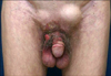
Physical examination revealed an oozing erythematous patch spreading over the pubic area, scrotum, and penis. Further, a 1×1 cm-sized solitary moist and fragile nodule was on the scrotum (Fig. 1). Inguinal lymphadenopathy was not evident. Biopsy specimens were obtained from the patch and the nodule.
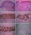
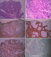
On microscopic examination, the patch showed large, round cells in the epidermis. Those cells were scattered discretely or grouped forming nests in the epidermis and showed pagetoid spreading (Fig. 2A). These pagetoid cells had large nucleus and pale cytoplasm, and intercellular bridges between the cells were not seen. The basal cells beneath these cells were somewhat flattened (Fig. 2B). Immunohistochemical stains for CEA, CK7, and CAM5.2 were expressed while the stains for CK20, HMB-45, S100, and P63 were completely negative (Fig. 2C~F). Otherwise the nodule showed irregularly thickened epidermis, which had cells lying in complete disorder, resulting in 'wind-blown' appearance (Fig. 3A). Many cells appeared highly atypical, showing large, hyperchromatic and pleomorphic nuclei and some of them showed atypical individual cell keratinization (Fig. 3B). Immunohistochemical stains presented CEA+, CK7+, CAM5.2+ and CK20-, HMB-45-, S100-, P63- (Fig. 3C~F). No other malignancy was found on workups, including radiologic examination. Finally, we diagnosed him as a primary EMPD with bowenoid features. He underwent total excisional surgery, and now he has been under observation with no recurrence so far.
DISCUSSION
In 1955, Woodruff1 suggested that the squamous epithelium and pilar apparatus, including apocrine and eccrine sweat ducts, were derived from the pluripotent embryonal germinativum. He told that EMPD derived from the malignant transformation of the basal stem cell that expressed gland differentiation1. Controversy in the histogenesis in EMPD and variability between reported cases seem to support the multipotent stem-cell theory2-4.
Now, a central feature to our understanding of EMPD is its association with gland-bearing skin and the immunohistochemical expression of glandular cell markers, like CEA, low molecular cytokeratines, and antibodies to GCDFP-155-7. In our case, histopathologic feature of the patch was consistent with EMPD. However, the nodule revealed a totally different feature, showing the characteristics of Bowen disease. However, the strong expression of CEA+, CK7+, CAM5.2+and P63- in both specimens made it possible to confirm the overall diagnosis of EMPD with bowenoid features. Bowen disease is negative for CEA, and it usually shows expressions of high molecular cytokeratins, but lacks of low molecular cytokeratins in a fashion similar to the normal epidermal keratinocytes8. Besides, Bowen disease expresses P63, which is essential for gulating the proliferation and differentiation of squamous cells9. Lack of CK20 expression and lack of other findings on laboratory and radiologic examination meant his EMPD was not from a pagetoid spread of extraneous malignancies, but from the primary.
There are several reports suggesting the association of squamous cell carcinoma in situ with EMPD8,10-14, Pagetoid Bowen disease, EMPD with bowenoid features, and EMPD admixed with Bowen disease, and so on. Like this case, those also support the multipotent stem-cell theory. In addition, thinking of the resemblance between EMPD and Bowen disease, dermatologists should avoid the pitfalls through appropriate immunohistochemical study when the diagnosis is ambiguous.
 XML Download