

 PDF
PDF ePub
ePub Citation
 Print
Print


INTRODUCTION
Genetic factors account for the majority of differences in skin color and hair morphology across human ethnic groups. Although many studies have been conducted to examine differences in skin color across populations, few studies have examined differences in hair morphology. However, interest in geographical variations in hair has increased in regards to the cosmetics industry. Previous studies confirmed both morphological and biochemical differences across ethnic groups. Morphological studies have focused mainly on hair color, shape, and responses to external stimuli, while biochemical studies have focused on the compositions of the hair proteins. Only a few studies have assessed the differences in hair lipids across groups. This may be due to the chemical composition of hair, which is composed mainly of keratin protein. Additionally, there are few established methods to examine lipids in human hair.
Hair lipids are composed of fatty acids, cholesterol sulfate, ceramides, and cholesterol. Together, these account for 0.7~1.3% of the total chemical content of hair1,2. Integral hair lipids are located in the cell membrane complex (CMC) of hair cuticles, and are important in the maintenance of hair integrity due to qualities including hydrophobicity, moisturization, and stiffness1,3. Integral lipids of the hair and epidermal lipids of the skin have similar barrier functions; however, little is known about the nature of population differences in the production of integral hair lipids1.
Forms of hair damage include physical stress, such as shampooing and combing, and chemical stress, like dyeing and bleaching. The most damaging among these factors is continuous exposure to sunlight. Ultraviolet (UV) light-induced hair damage is difficult to avoid4,5. Continuous UV-light exposure results in dryness, roughness, sun-bleaching and breakage due to photo-oxidation6.
In this study, we examined integral hair lipids in three human populations. The results of UV-induced integral hair lipids differed across groups.
MATERIALS AND METHODS
Materials
Standardized (age and sex matched) hair samples from individuals originating from three different geographical ethnic groups (Asian, European, and African) were purchased from De Meo Brothers, New York, NY, USA. The hair samples (hair tresses) were washed with a 3% diluted commercial shampoo, and then rinsed thoroughly with water and dried in ambient conditions7. Since there are some limitations for sampling of the hairs in regard to each race, we used the commercial samples.
UV irradiation
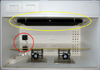
UV irradiation was performed using a phototherapy system composed of six perpendicular UVA lamps (20 W, FL20SBLB, Sankyo Co., Ltd., Tokyo, Japan) and UVB (8 W, G8TE, Sankyo) (Fig. 1). Damaged hair samples were prepared at 12 and 48 hours after UVA (20 J for 12 hours and 40 J for 24 hours) and UVB (8 J for 12 hours and 16 J for 24 hours) irradiation. The distance from the lamp to the hair samples was 50 cm, and the relative humidity was 30%. A cooling fan was used to prevent the heat damage due to UV irradiation. All experiments were conducted at room temperature7.
Ultrastructural findings
1) Scanning electron microscopy (SEM)
Each prepared hair sample was affixed to a specimen slide, and gold coating was applied to the fixed hair sample with a sputter coating machine. Slides were examined by SEM (LEO 1499AP, 30 kV, LEO, Germany). Results were scored according to a SEM standard grading system, as previously described by Kim et al.7.
2) Transmission electron microscopy (TEM)
All hair samples were incubated in 1:1 mixture of propylene oxide and epon overnight and then embedded in an epon mixture. Horizontal sections of approximately 60~70 nm were prepared and stained with uranyl acetate and lead citrate. The specimens were viewed under TEM (JEM-1200EDXII, 80 kV, JEOL, Tokyo, Japan)8. The results were scored according to the TEM standard grading system7.
Integral hair lipid analysis
Twenty milligrams of each hair sample were cut into 4 cm lengths, and continuously immersed in 5 ml of chloroform-methanol (2:1, 1:1, and 1:2) for 24 hours. The last immersion was in a chloroform-methanol-water (18:9:1) solution for 24 hours. The extracted solutions were pooled and filtered through a 0.5 µm Millipore filter. The filtrates were united and dried under a nitrogen stream, and the residue was recovered by dissolving the filtrates in 100 ul of the 1:1 chloroform-methanol mixture for further HP-TLC analysis. The integral hair lipids were dissolved in 100 ul of the 1:1 chloroform-methanol mixture, and 2 ul of each solution containing lipids were spotted onto silica gel-coated rods using a microdispenser. The rods were developed three times consecutively using the following: (i) hexane-diethyl ether-acetic acid (200:80:1) up to 6 cm; (ii) hexane-benzene (1:1) up to 8 cm; and (iii) hexane up to 10 cm. After each development, the rods were heated at 60℃ for 5 minutes in order to dry. After completion, the rods were treated with diluted H2SO4, and were heated at 180℃ for approximately 15 minutes.
RESULTS
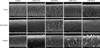
Evaluation of hair surface damage by SEM
We evaluated the damage to the hair surface due to UV irradiation, using a standard grading system for damaged hair7. The hair cuticles from all three groups had intact and tightly overlapping cuticle scales. As the irradiation energy increased, the surfaces of samples from all three groups became increasingly damaged. Focal lift, loss of the cuticle edge, and exposure of the cortical cells were observed in all samples. When the grading system was applied, the grading scores for each group increased as the exposure to UV irradiation increased (Table 1). UVB irradiation resulted in more severe damage than UVA irradiation, and compared to the samples from other groups, the African hair exhibited relatively worse damage (Fig. 2).
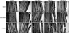
Evaluation of cuticle layer damage by TEM
All hair samples across groups had intact cuticles of greater than six layers. As the irradiation energy increased, variably sized holes, cleavage along the endocuticles, and cuticular detachment were observed in all groups. Consistent with the SEM results, the grading score increased with increased UV irradiation exposure (Table 2). UVA irradiation and UVB irradiation resulted in similar damage. The hair samples of all three groups exhibited similar patterns of damage. However, the African hair exhibited the weakest resistance to UV irradiation (Fig. 3).
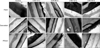
Evaluation of lipid layer damage by lipid TEM
All hair samples had intact intercellular lipid layers prior to UV irradiation. Following UV irradiation, we observed some focal bulging and disruption in the intercellular lipid layers. UVA and UVB irradiation resulted in similar changes to the intercellular lipid layers. UVA irradiation resulted in swelling of the lipid layer, and UVB irradiation caused disruptions to the lipid layer. We confirmed that the more energy exposure to UV radiation, the greater the damage to the lipid layer. In particular, African hair exhibited relatively weaker resistance to the UV irradiation than the samples from other groups (Fig. 4).
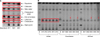
Integral hair lipid analysis by HP-TLC
Among the samples of the three groups, differences were found in the integral hair lipids. Asian samples had more integral hair lipids than others prior to UV radiation exposure (Fig. 5A) and after exposure (Fig. 5B). Free fatty acid contents were higher than other lipids. Squalene content was higher in the African samples than in the other groups. UV irradiation decreased the lipid content, especially the free fatty acid content in the European and African samples. In the European and African samples, the levels of free fatty acids decreased when UV irradiation was performed. However, the Asian samples showed no change (Fig. 5).
DISCUSSION
The curliness, color, and cross-sectional parameters of hair samples differ across human ethnic groups. However, some proteins, keratins, and molecular structures, including amino acids, have common features across groups9-11. Although there are no significant geographical differences in keratin proteins, which are the main structural components of hair, physical characteristics differ among groups. Other components, such as melanin and lipid components, affect the characteristics of hair. Franbourg et al.9 reported that African hair exhibited less radial swelling when flushed with water compared to Asian or European hair and the authors assumed that there might be differences in hair lipids among human populations. However, proper lipid analysis of this study was not performed. Additionally, Syed et al.12 reported differences in moisturizing rates associated with the differences in lipid content. In hair care, many experts recognize the importance of moisturization; however, it is difficult to observe integral hair lipids and distinguish them from the lipids produced by sebaceous glands.
The lipid layer is important for maintaining hair integrity; thus, it is also referred to as the "hair barrier". During the process of keratinization in the cortex, an adhesive layer (CMC) forms between adjacent cells through the cortical plasma membrane. CMC is a continuous structure composed of three-layered structures, and the middle layer (δ-layer) is surrounded by inner and outer β-layers. The integral hair lipid is located in the beta-layer13. The lipid layer of hair is composed mainly of fatty acids, cholesterol sulfate, ceramide, and cholesterol. Orwin14 observed that globular particles in the intercellular spaces form lamellar structures during hair development. When they migrate to the external area and membrane rupture is occurred, these particles form the lamellar structure. Finally, a lipid layer builds up between the outside of the hair cuticle and the IRS cuticle. One of the most important lipid components is 18-methyleicosanoic acid (18-MEA), which occupies roughly 40% of all fatty acids and has an ester bond or thioester bond with the keratinized cell15. CMC is a major component of 18-MEA, and it is the lipid layer that decreases friction and increases hydrophobicity of the hair surface13. We determined that samples from Asians demonstrate higher levels of integral hair lipids than other groups. Free fatty acids, cholesterols, and wax esters were also higher in Asian samples. African samples exhibited higher levels of squalene, but overall, lipid content was lower for this group than for Europeans or Asians. Previous reports9,12 using hair swelling revealed that hair lipids of Africans may be more prominent than other ethnic groups. Both our results and previous results may be contradictory to one another. Previously reported results were made from non-standardized hair samples and it may cause the cause for the different results. We used standardized hair samples but there are many factors which can affect the lipid contents that cannot be determined. To analyze more accurate lipid contents, further advanced studies are needed.
UV light is composed of UVA (320~400 nm), UVB (290~320 nm), and UVC (200~290 nm) waves. UVC is blocked by the ozone layer, but UVA and UVB affect skin and hair. Damage to hair due to UV light occurs due to free radicals or cysteic acid, which forms after UV radiation and breaks disulfide bonds16,17.
In this study, SEM and TEM analyses showed similar patterns of hair damage, regardless of population origin. As UV irradiation increased, greater focal lifting and loss of the cuticle edge was observed. Furthermore, the number of cuticle layers decreased after 48 hours of UV irradiation. We confirmed that hair surfaces are damaged primarily by UVB light, while hair lipids are damaged primarily by UVA light. These results may be due to differences in the penetration depth of UV light4.
Damage to the integral lipid layer was similar across groups. Normal hair had a uniform lipid layer, but UV damaged hair displayed swelling of the lipid layer and disruption. The bulging of the lipid layer was mainly observed in samples exposed to UVA irradiation, and the disruption of the continual lipid layer was mainly observed in samples exposed to UVB irradiation.
Lipid content was higher in Asian samples than in other groups, while free fatty acid levels were lower in African samples than in other groups. After UV irradiation, the Asian samples were less damaged than the African samples, while the damage to the European and African samples was time dependent. Thus, we inferred that the quantity of free fatty acid is likely associated with hair damage though there are several additional factors that might also account for the observed effect, such as fiber geometry, diameter and pigment content which need to be investigated from this point forward.
In conclusion, we compared and analyzed the integral hair lipids across three populations and observed morphological changes of hair lipids following UV irradiation. We determined that Asian hair contains more integral hair lipids than European or African hair. After UV irradiation, African and European hair samples exhibited more damage than Asian samples. Therefore, we concluded that integral hair lipids may protect hair against UV light.


 XML Download