

 PDF
PDF ePub
ePub Citation
 Print
Print


Dear Editor:
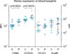
Chronic urticaria (CU) is currently diagnosed clinically when patients weals and the surrounding reflex erythema persist for more than 6 weeks1. Autoreactivity of the CU-patients sera can be established in 30% to 50% of patients, depending on the population, through the autologous serum skin test (ASST)2, the basophil activation test based on upregulation of CD63 (BAT)3 or basophil histamine release assay (HR), the latter which is now available as a commercialized testing kit in the market. The ASST has only moderate specificity as a marker for functional autoantibodies against the high-affinity immunoglobulin E (IgE)-receptor FcεRIα2. Recent research on the field has shown significant upregulation of activation markers CD63 and CD203c on whole blood basophils from CU-patients as well as an up-regulation of expression of the high-affinity IgE-receptor FcεRIα4. There is a need for a robust laboratory test for the diagnosis of CU, which can persistently and objectively support the anamnestic diagnosis1. The purpose of this study was to confirm the measurement of in vivo expression of the activation markers CD63, CD203c and CD123 (interleukin-3 Rα receptor) and the high-affinity IgE-receptor FcεRIα using flow cytometry4 as an objective complement to anamnestic diagnosis of CU. The study included 8 patients diagnosed with CU, according to the EACCI/GA(2) LEN/EDF/WAO guidelines1, (mean age 43 years; positive HR test, n=2; negative HR test, n=3; HR test not performed, n=3) and 12 healthy control subjects with a mean age of 48 years. Within 3 hours after venipuncture, 100 µl aliquots of EDTA blood were stained at room temperature for 30 minutes with CD123 PE (BD Pharmingen 340545, 10 µl) and FceRI (Biolegend 334614, 1 µl) in tube 1, CD63 FITC (Biolegend 312004, 5 µl) and CD193 Alexa 647 (Biolegend 310710, 2.5 µl) basophils in tube 2 and CD203c (IOTest PMIM 3575, 5 µl) and CD193 Alexa 647 in tube 3. Samples were hemolyzed and washed with phosphate buffered saline. Data was acquired on a FACS Canto II (Beckton-Dickinson, San Jose, CA, USA) flow cytometer for 4 minutes at medium speed in order to obtain absolute cell numbers from the constant volume sampled5. Performance of the flow cytometer was monitored with Cytometer Setup and Tracking beads, and an external quality assurance program. Basophil granulocytes were identified as FceRI+ or CD193+ cells with low side scatter. Patients and controls were compared with the Mann Whitney U test. In line with previous studies6,7, the CU patients within this study had pronounced basopenia (median 358, interquartile range [IQR] 145 to 413) vs. 669 (IQR 476 to 891), p<0.002). CU-patients' basophils expressed significantly more CD63 and CD203c (both p<0.003, Fig. 1). There was no difference in the expression of FceRI or CD123. It is possible to set thresholds to classify patients as likely to have urticaria on the basis of blood basophil concentration of 431 cells/4 minutes, basal CD63 median fluorescence intensity (MFI) > 1,636 and CD203c MFI > 2,160 with area under the curve of 0.92, 0.88 and 0.90, respectively. Our findings correlate with the findings of Lourenço et al.4 regarding the basopenia of CU-patients and upregulation of CD63 and CD203c; however, we did not find an increase in FcεRIα-expression. In a similar study on enriched basophil suspensions, CD203c expression was not found to be up-regulated with urticarial subjects7. This may be because even a partial purification of basophils can profoundly limit the basophil function8. With our small dataset, it was not possible to determine a correlation of high expression of activation markers CD63 and CD203c with positive HR. The patients in our study were seen on an out-patient basis and thus, it is not possible to conclude from our study for how long the baseline activation markers are raised. Further investigations into baseline basophils activation markers in direct timely relation to disease outburst and attack are needed. Our study does however contribute with evidence of the activated profile of basophils in CU. We confirm that a flow cytometric analysis of basophil concentration as well as basal expression of CD63 and CD203c in patient blood may be used to diagnose patients with CU. This may then be investigated for the involvement of autoantibodies by a basophil activation test. Further studies are needed to investigate if CD63 and CD203c may be used to monitor the efficacy of treatment. Furthermore, our findings indicate that the activation of basophils may be an important event in the pathogenesis of CU.
 XML Download