

 PDF
PDF ePub
ePub Citation
 Print
Print


Dear Editor:
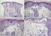
Focal acantholytic dyskeratosis (FAD) was first described by Ackerman1 in 1972 as a distinct histopathological pattern associated with various cutaneous conditions, and with classic histopathological findings including suprabasal clefting, hyperkeratosis and parakeratosis, and the presence of acantholytic and dyskeratotic cells at the epidermis. While FAD can be observed in many various cutaneous lesions including benign and/or malignant epithelial lesions, fibrohistiocytic lesions, inflammatory lesions, melanocytic and/or follicular lesions2-4. These histopathological findings may also extend into the surrounding tissues, which often appear to be clinically normal. A 42-year-old woman was presented to our department with multiple erythematous pruritic papules and tiny vesicles on her face. The lesions had been present for several years and aggravated 7 days ago. Physical examinations revealed multiple 2 to 3 mm, slightly spongiotic-appearing papules and tiny vesicles with serous crusts on the face (Fig. 1). Laboratory tests obtained at that time were within normal limits. A skin biopsy of an erythematous papule on the nose was also performed, and histopathological results revealed focal suprabasilar clefting and acantholytic keratinocytes in the epidermis, dense inflammatory infiltrates and vascular dilatation with solar elastosis in the dermis, and a negative direct immunofluorescence (Fig. 2). In the serial sections, we observe the same findings. Differential diagnosis for the possibility of polymorphous light eruption, systemic lupus erythematosus, contact dermatitis, and dermatitis artefacta should be considered. But given these clinical and histopathological features, a diagnosis of rosacea with FAD was reached. The patient was then admitted to hospital for treatment with doxycycline 100 mg and antihistamines. After one week, the lesions had remarkably improved. The patient was then discharged, and continued on the same therapeutic regimen for an additional month, bythe time all lesions were nearly resolved.
To date, the etiology of FAD has been attributed to numerous sources including hormones, viral infection, various immunologic factors, tobacco use, physical stimuli although the exact causative mechanism of this finding remains unknown. Other researches have suggested that sunlight and/or ultraviolet radiation may lead to the development of FAD5.
In our case, we propose that the acantholytic dyskeratosis occurred secondarily to ultraviolet radiation exposure, given the lesion's location on the nose, a chronically sun-exposed area. Furthermore, chronic physical irritations may also have influenced, as the patient complained of severe pruritus limited to the affected area, the resulting excoriations which possibly lead to acantholytic dyskeratosis.
To the best of our knowledge, there have not been any prior reports of FAD associated with rosacea. We also contended that UV exposures combined with consistent physical irritation (i.e. excoriation) which represent two prime etiological factors contributing to the development of FAD in our patient. Therefore we report herein a case of FAD associated with rosacea and this report may provide additional explanation of pathomechanism in incidental FAD in the setting of rosacea.

 XML Download