

 PDF
PDF ePub
ePub Citation
 Print
Print


INTRODUCTION
Topical glucocorticoid therapy is one of the most commonly used immunosuppressive or anti-inflammatory drugs. Even with the invaluable therapeutic efficacies, however, inevitable local side effects including skin atrophy, striae, telangiectasis, prupura, acneform eruption, perioral dermatitis, and steroid-induced rosacea impede the long-term use of steroids for chronic skin diseases such as atopic dermatitis. Recently, it was also reported that both the long-term and short-term application of topical steroids can compromise the skin barrier function, mainly by inhibiting the epidermal lipid synthesis1,2.
Various strategies to minimize the steroid-induced side effects have been suggested, including modification of application schedules3-5, designing new steroid molecules6, and adjunctive use of other drugs with anti-atrophogenic activity7. Use of new vehicles for topical steroids is also suggested as an effective way of reducing the side-effects of steroids, as well as improving the transcutaneous delivery and epidermal release profile. It has been reported that a hydrogel formulation containing desonide, a low potency corticosteroid, had an improved risk-benefit ratio in atopic dermatitis patients8,9. Recently, we have shown that a multi-lamellar emulsion (MLE), as a vehicle for clobetasol 17-propionate, is effective for preventing the steroid-induced skin atrophy and epidermal permeability barrier function impairment10.
In the present study, the effects of MLE as a vehicle for topical steroids is further investigated. Desonide, a low potency glucocorticoid, is formulated into MLE, and the anti-inflammatory activity and transcutaneous delivery were measured. Steroid-induced side effects on skin atrophy and skin barrier function were also evaluated in animal model.
MATERIALS AND METHODS
Materials
Stearic acid, cholesterol, and fatty acid triglyceride were purchased from Junsei Chemical Co. (Tokyo, Japan); and glyceryl monostearate and POE(15) glyceryl monostearate were from Nihon Emulsion Co (Tokyo, Japan). Cetanol (Kao Co., Tokyo, Japan) and squalane (Kishimoto Co., Hyogo, Japan) were used for the preparation of emulsions. Synthesis of pseudoceramide (myristyl/palmityl oxostearamide/arachamide MEA: C34H67NO3/C36H71NO3/C38H75NO3=11 : 42:47%) and its chemical characteristics were described in a previous report11. Desonide was obtained from Hubei Gedian Humanwell Pharmaceutical Co., Ltd. (Hubei, China). Commercial cream and lotion products containing same 0.05% desonide were used for comparison.
Preparation of test formulations
In order to prepare MLE vehicle, the lipid components (pseudoceramide, stearic acid, and cholesterol), oils, common base (cetanol, 2-octyldodecanol, squalane, and fatty acid triglyceride) and emulsifiers (glyceryl monostearate, POE(15) glyceryl monostearate) were mixed and melted at 85 to 90℃. The desonide was dissolved in 1, 3-butylene glycol and then slowly added with vigorous agitation by homomixer (T.K. Homomixer Mark II f-model, Tokyo Co., Ltd., Tokyo, Japan) at 70℃. After emulsifying, the mixture was cooled down to the room temperature. All formulations were prepared containing 0.05% desonide of the final content (Table 1). Detailed preparation scheme was previously reported12.
In vivo efficacy measurement
Female hairless mouse (8 weeks old) and male ICR mice (4 weeks old) were purchased from Orient Bio Co. (Seongnam, Korea) and kept under controlled humidity (50~60%) and temperature (23~25℃) with a 12 hr/12 hr (light/dark) cycle. All the animal experiments described were approved by the Institutional Review Board of Yonsei University College of Medicine.
In order to observe the steroid-induced skin atrophy, tested formulations were applied to the entire dorsal skin of hairless mouse twice a day for 3 days. Skin double-fold thickness was measured using digital calipers (Mitutoyo Co., Ltd., Kawasaki, Japan). Epidermal permeability barrier function and skin hydration were evaluated by measuring trans-epidermal water loss (TEWL) and skin capacitance, respectively. Measurement of TEWL and skin capacitance was performed using Tewameter TM 300 (Courage & Khazaka, Cologne, Germany) and Corneometer CM 820 (Courage & Khazaka), respectively.
Anti-inflammatory activity of desonide formulation was evaluated using mice ear model, and the skin inflammation was induced by repeated treatment of 12-O-tetradecanoylphorbol-13-acetate (TPA) for 7 days. TPA (1 µg/20 µl acetone) was applied to the mouse ear (day 0). The tested formulations were then topically applied to the same site (20 µl) at 1 and 9 hours after TPA treatment (day 1~day 6). At day 7, tested formulations were applied at 1 hour after TPA treatment; and after 3 hours, skin biopsy was taken for further analysis. Modulation of inflammatory marker mRNA expression was measured by real-time reverse-transcriptase (RT) polymerase chain reaction (PCR). Mouse ear samples were homogenized in buffer RLT (lysis) buffer for 2 minutes using T-10 ultra-turax homogenizer (Staufen, Germany). Total RNA was isolated using RNeasy mini kit (Qiagen, Valencia, CA, USA), and corresponding cDNAs were synthesized using RT reaction at 4℃ for 1 hour and 95℃ for 5 minutes. Primers were synthesized for interlukin-1β (IL-1β) and tumor necrosis factor (TNF)-α, and GAPDH. The primer sequences used for real time-PCR were as follows: IL-1β sense, 5'-CCC AGC AAT ACC CAA AGA A-3', antisense, 5'-GCT TGT GCT CTG CTT GTG AG-3'; TNF-α sense, 5'-AGC CCC CAG TCT GTA TCC TT-3', antisense 5'-GGT CAC TGT CCC AGC ATC TT-3; GAPDH sense, 5'-ATG ACA TCA AGA AGG TGG TG-3', antisense, 5'-CAT ACC AGG AAA TGA GCT TG-3'. Real time-PCR was carried out for 55 cycles in 10 µl reaction mixture. PCR reaction hold was at 95℃ for 15 minutes, denaturing at 95℃ for 10 seconds, annealing at 57℃ for 15 seconds, and extension at 72℃ for 20 seconds in Rotor gene-6000 (Corbett Life Science, Sydney, Australia).
Histological assessment was performed by conventional haematoxylin and eosin (H&E) staining and immunohistochemical staining. Biospy samples were fixed with 10% formalin and embedded in paraffin. Four micrometer thick sections were cut and stained with H&E staining, and observed under microscope.
Penetration experiments
Female hairless mouse were sacrificed with cervical dislocation, and full-thickness abdominal and dorsal skin of mice were excised. The extraneous subcutaneous fat was removed from the dermal side, and the skin samples were stored in freezer at -20℃ and used within 2 weeks. Immediately before use, skin samples were thawed and hydrated in 0.05% sodium chloride solution for 30 minutes and cut into appropriate pieces. The skin sections were mounted on surface areas of 0.785 cm2 occluded Franz diffusion cells and the acceptor compartment was filled with phosphate buffer pH 7.4. The epidermal side of the skin was exposed to ambient environment and allowed to equilibrate for 1 hour before experiment. After equilibration, 40 mg of each formulation was placed on skin surface using a spatula and receptor solution was maintained at 37±0.5℃ and stirred continuously at 600 rpm. The receptor solution (4 ml) was taken at predetermined times over 24 hour, and the volume was replenished immediately with buffer solution. The experiments were carried out in triplicate.
Determination of the residual desonide in skin
After 6 hour of permeation sampling, the skins were removed from the diffusion cells and washed with distilled water. Then, the skins were punched using 6 mmdiameter punch and immersed in 10 mM EDTA-PBS solution overnight at 4℃. The epidermis of the skins was then peeled off carefully with a blunt forceps. The epidermis and dermis samples were desiccated for 24 hour and weighed. The samples were put in a 5 ml tube and cut in small pieces, and 1 ml of methanol was added. After that, the samples were homogenized ten times for about 30 seconds using ultrasonic processor (VCX500, Sonics and Materials Inc., New Town, CT, USA). The liquid phase was centrifuged at 10,000 g for 10 minutes at 4℃. The upper phase was recovered and filtered using a 0.45 µm filter, and desonide concentration was analyzed using high performance liquid chromatography (HPLC).
Analysis of samples was performed by reverse phase HPLC composed of an auto sampler (Waters 717, Waters, Milford, MA, USA), a pump (Waters 600S, Waters), an photodiode array detector (Waters 996, Waters) and a column (Capcell Pak C18 UG120 5 µm 4.6×250 mm, Shiseido Fine Chemicals, Tokyo, Japan). The mobile phase (acetonitrile : distilled water, 4 : 6) was eluted at a flow rate of 1.0 ml/min, and the ultraviolet detector was set to 254 nm wavelength. The volume of sample injected was 50 µl. At this condition, the retention time of desonide was 7.2 minutes, and the minimal amount detected was 0.025 µg/ml. Desonide was quantitated using an external standard technique. Because of the low concentration of desonide in the skin tissues, all of the samples obtained in triplicate experiments were collected together and analyzed after concentrating the samples.
Statistical analysis
All results were represented as arithmetic mean±standard deviation. In order to evaluate the statistical significance, one-way ANOVA with post-t-test was performed using GraphPad Instat version 3.10 (GraphPad Instat Software, San Diego, CA, USA), and p-value less than 0.05 was considered as significant.
RESULTS
Therapeutic activity of desonide formulations
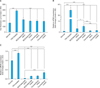
Chronic skin inflammation in ICR mouse ear was induced by repeated application of TPA for 7 days, including sensitization period. Significant hyperplasia and edema were observed in TPA treated site, and topical treatment of desonide containing formulations significantly reduced hyperplasia. Reduction of skin thickness was similar for all the tested formulations, compared to the TPA-only treated site (Fig. 1A). The epidermal expression of inflammatory cytokines, IL-1β, and TNF-α also significantly decreased in desonide-treated skin (Fig. 1B, C), which suggested that MLE as a vehicle did not affect the anti-inflammatory activity of desonide. Interestingly, lotion formulations were more effective for both the epidermal thickness reduction and inflammatory cytokines reduction than cream formulations in all the tested formulations.
Effects of MLE on the steroid-induced side effects
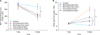
One of the most characteristic side effects of topical steroids is skin atrophy. In order to evaluate the skin thinning effects, tested formulations were topically applied on normal hairless mouse skin twice a day for 3 days. As a result, similar reduction of skin fold thickness was observed in all the tested formulations (Fig. 2A). Impairment of epidermal permeability barrier function, another important side effect of topical steroid, however, was less severe in MLE formulation. After 3 days application, TEWL, as a representation of epidermal permeability barrier function, significantly increased for all the tested formulations. While the commercial desonide products treated site showed statistically significant increase in TEWL value, MLE based desonide products showed a slight increase in TEWL, which was not statistically significant (Fig. 2B) compared to control group. In previous reports, Kao et al.2 showed that topical treatment of glucocorticoid induced an impairment of epidermal permeability barrier function, which was, at least in part, mediated by inhibition of epidermal lipids synthesis. It was also suggested that topical supplement of these stratum corneum intercellular lipids can reduce the steroid-induced side effects. Previously, we also reported that topical application of high potency glucocorticoid, clobetasol-17-propionate with physiologic lipids mixture can reduce the steroid-induced side effects, which is consistent with present study10.
In vitro skin permeation and accumulation study
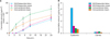
Since the penetration of topical steroid plays a crucial role in its therapeutic efficacy and side effects, the transcutaneous penetration of steroid and its skin accumulation were evaluated using excised hairless mice skin. Mea surement of TEWL before and after penetration study confirmed an intact permeability barrier function for the used skin tissues (data not shown). While the commercial desonide products showed similar penetration of desonide at the earliest sampling time (2 hours after application), MLE/desonide cream and lotion took about 4 hours to be detected in the receptor fluid, which suggested that the MLE formulations were less efficient for transcutaneous drug delivery for topical steroid (Fig. 3A). However, the major site-of-action for topical steroid is epidermis and dermis, and we measured the residual amount of desonide in epidermis and dermis after 6 hours application. While the residual amounts in dermis were nearly comparable for all the tested formulations, epidermal concentration was significantly different. While the lotion-based formulations showed higher concentration than cream-base for both formulations, MLE/desonide lotion showed more than 4-fold increased concentration than other formulations (Fig. 3B).
DISCUSSION
Desonide is a synthetic, non-fluorinated low-potency glucocorticoid, which is most commonly prescribed for atopic dermatitis by the dermatologist in the USA. With its lower incidence of local and systemic adverse effects, wide range of topical formulations from classical ointments, creams, and lotions to the recently introduced gel and foam further improved the patient's compliance to topical desonide treatment. In addition to the role of carrier and stabilizer for efficient and effective drug delivery, vehicle for topical therapies can also influence the patient compliance. In this study, we have evaluated the effects of MLE as a vehicle for topical desonide therapy.
In addition to the well-known local adverse effects including skin atrophy, impairment of epidermal permeability barrier function and epidermal anti-microbial peptides (AMPs) expression are recently suggested as important adverse effects of topical glucocorticoids. Previous studies established the critical role of stratum corneum intercellular lipid lamellae in epidermal permeability barrier function. Recently, it was reported that long-term application of topical steroid induced the structural defects in intercellular lamellae structures, as well as impaired skin barrier function1. Interestingly, even short-term application of topical steroid also induced skin barrier function impairment, due to the inhibition of epidermal lipid synthesis and disruption of coreneodesmosome. Simultaneous application of physiologic lipid mixtures, which composes stratum corneum intercellular lipid lamellae, significantly reduced the steroid induced barrier function impairment2. These reports suggest that, unlike atrophogenicity, skin barrier function impairment can be effectively prevented by additional supplementation of physiologic lipid components. We have reported synthesis of novel ceramide derivatives as a structural analogue for natural ceramide in human stratum corneum13. In a series of works using the newly synthesized ceramide derivatives, the formulation of MLE, characterization of its structural property, and its clinical benefits on skin barrier function were reported11,12,14. In this study, while the reduction of skin thickness was similar for all the tested formulations, skin barrier impairment by topical desonide was nearly completely prevented in MLE-based desonide formulations, which is consistent with previous studies. Since various steroid-responsive dermatoses are known to show the impairments of skin barrier functions, the beneficial effects of MLE on skin barrier functions may improve the clinical outcomes of topical steroids treatments.
Another important barrier function of skin is the antimicrobial barrier, which is mainly exerted by AMPs15. AMPs are generally small cationic polypeptides having an anti-microbial activity, as well as modifying inflammatory reactions and immune reactions. Recently, reduction of AMPs in lesional skin of atopic eczematous patients was reported, which was considered as the underlying mechanism for an increased microbial susceptibility of atopic skin16. Among the various factors affecting AMPs expression, glucocorticoids are well-known suppression factors17,18. In present study, reduction of AMPs was observed in steroid applied skin, and preventive effect of MLE was also observed. However, the differences between normal skin and steroid-applied skins were not significant, possibly due to the low potency of used steroids. Using a high potency steroids, additional studies need to be performed to verify the effects of topical steroid on epidermal AMPs expressions and the preventive effects of MLE on AMPs reduction.
Measuring the transcutaneous penetration of desonide in this study resulted in a higher transdermal drug delivery of the lotion formulations compared with cream preparations. Among the diverse factors affecting the drug delivery, differences in ingredients or ingredient's concentrations may have rendered the skin more permeable to steroid for lotion formulations. Various ingredients were used for preparing the vehicles, and some ingredients were known to have a penetration enhancing effect, or a penetration retarding effect. Complex interaction of these ingredients' effects and interaction with skin also resulted in a difference in steroid penetration between cream and lotion formulations. Comparing the conventional formulation with MLE formulations, much higher effectiveness for penetration was observed in conventional formulations. The site-of-action for topical steroids, however, is viable epidermis and dermis; and as such, the residual amount of steroid in skin is more important than penetrated amount. Interestingly, measuring the residual amount of desonide in epidermis and dermis showed that significantly higher amount of desonide was accumulated in epidermis, compared with all the other tested formulations. Since the MLE formulations were prepared for mimicking the native structure of human stratum corneum intercellular lipids, structural similarity of MLE formulations might increase the retentions of the topically applied drugs in skin, especially in epidermis. However, more investigations need to be performed to explain the differences between MLE lotion and MLE cream formulations.
While the topical glucocorticoids are one of the most important therapeutic regimens for atopic eczema, more patients are becoming reluctant to use topical steroids due to their significant side-effects. Even with enormous efforts to overcome the side-effects, it still remains to be further investigated for more safe steroid formulations. Recently, development of novel hydrogel vehicle for topical steroid was reported, and it was shown to be more effective for skin moisturization and skin barrier function9. In this report, we also showed that use of MLE can prevent the impairment of skin barrier function. In addition, transcutaneous delivery profile was more preferable in MLE formulation, especially in MLE lotion formulations. Since the therapeutic anti-inflammatory activity of steroid was not affected by the use of MLE vehicles, these results suggest that MLE as a vehicle for topical steroid can prevent steroid-induced side effects and improve patient compliance to the therapy, especially in atopic eczematous patient.

 XML Download