

 PDF
PDF ePub
ePub Citation
 Print
Print


INTRODUCTION
A common etiologic background may be inferred from the coexistence of two or more autoimmune associated diseases. Vitiligo is an unknown disease in which genetic and neurohumoral factors are thought to be causative agents besides autoimmunity. Etiology of Lichen planus is also unknown and immune factors are thought to play a key role in the pathogenesis. Vitiligo and lichen planus both occur concomitantly with other autoimmune diseases. Regarding a relative prevalence of 0.5~1% of lichen planus and vitiligo, coincidence of these diseases in one patient can be predictable, but development of these two unrelated diseases in the same anatomic areas is unusual, rare and has been described by only few reports1,2 in the last ten years. The occurrence of both conditions in one patient seems to not be just an accidental phenomenon as a probability of autoimmune background or common pathogenesis exists. Here, we report a very rare colocalization of lichen planus and vitiligo in two members of a family. To our knowledge, no other such cases have been reported.
CASE REPORT
Case 1: mother
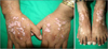
The mother is a 37 year old lady, farmer with a known case of vitiligo from 7 years prior. She presented with a one year history of pruritic skin eruptions over here hands and feet. Physical examination revealed multiple well defined depigmented patches over feet, hands, trunk, elbows and knees with multiple pinkish to erythematous plaques predominantly localized over hands and feet confined to sun exposed and hypopigmented vitiliginous areas (Fig. 1). Her elder sister also had pathologically proven lichen planus lesions over her face and hands without vitiliginous patches.
Case 2: daughter
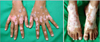
The daughter is a 23 year old single woman, farmer with a known case of vitiligo for 10 years, presented with a history of pruritic skin eruptions that begun over her hands and feet, and extended to her forearm and face since 6 months earlier. Physical examination showed multiple hypopigmented vitiliginous patches over the hands, feet, posterior of thighs, knees, and arms with multiple pinkish to erythematous plaques over hands and feet. The patches were present over sun-exposed areas confined to depigmented vitiliginous areas with gradual violacious discoloration when extending to normally pigmented skin. There were also few violacious to brownish macules and plaques over the forearm, face and lips (Fig. 2).
In both patients oral mucosa and nails were normal and preceding vitiliginous areas including knees, trunk, legs and forearms were free of the new lichen skin lesions. Paraclinical and laboratory examinations including biochemistry tests (blood sugar, blood urea nitrogen, creatinine, electrolytes, liver enzymes, alkaline phosphatase, and bilirubin), the thyroid function test and serology tests of C3, C4, immunoglobulin (Ig)G, IgM, IgA, IgE, hepatitis C virus antibody, hepatitis B-surface antibody, antithyroid peroxidase antibody were all within normal limits except for the high level of IgE in daughter which was 435.90 KIU/L (normal range was up to 150).
The skin biopsy of lesions on hands confirmed lichen planus in both patients, showing hyperkeratosis, acanthosis, hypergranulosis, and basal cell liquefaction with civatte bodies associated with band like infiltration of chronic inflammatory cells in the dermis.
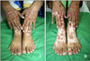
On the basis of the clinical and histological diagnosis of lichen planus, systemic and topical steroids were initiated. Marked improvement was achieved after one month resulting in lichen planus skin lesions with post inflammatory hyperpigmentation in non-vitiliginous areas and repigmentation of some previous vitiliginous areas as well (Fig. 3).
DISCUSSION
Immunity plays a direct role in the etiology of lichen planus and vitiligo. Coincidence of these two skin diseases has been reported exclusively or concomitant with other autoimmune diseases1,2.
Anstey and Marks3 proposed the presence and interference of lymphocytes against dermo-epidermal junction antigens in the development of vitiligo and lichen planus in one patient and concluded that immunogenic mechanisms and nonspecific deactivation of immunologic reactions cause the occurrence of these two diseases in one patient.
The occurrence of both psoriasis and lichen planus is also reported in a case of vitiligo. A reduced amount of melanin in one area could be an underlying cause for the development of other diseases in the same area. Lichen planus and psoriasis both have a positive koebner phenomenon. It seems that cell damage in vitiliginous areas incites immunity reactions and the koebner phenomenon. Ujiie et al.4 also referred to this topic and suggested that the koebner phenomenon and sun exposure can be effective in initiating psoriasis, lichen planus and vitiligo.
In our cases, the presence of lichen planus lesions in sun exposed areas indicates that sun rays are a causative agent in the occurrence of new lesions. Constellation of lichen planus lesions in depigmented areas especially in hands and feet over vitiliginous lesions demonstrates that the absence of melanin plays a significant role in inflammatory reactions following sun exposure. In our patients, lichen planus lesions are less severe on normal skin, and absent especially on vitiliginous lesions in covered areas such as the knees andelbows. In other words, perhaps depigmentated areas play a key role in inciting lichen planus. Lichen planus begins from depigmentated areas and then extends to normal skin.
Usually generalized, familial lichen planus starts at an earlier age in children and young people, which can be prolonged with a higher recurrence rate. Familial vitiligo is a disease primarily found in the elderly as well, having a prolonged course with more associated concomitant diseases. Familial incidence of lichen planus and vitiligo in two family members makes the importance of genetic factors obvious in the pathogenesis of these diseases.
The probability of inheritance in first degree relatives was reported to be approximately 56% in lichen planus5 and the frequency of vitiligo in probands' siblings was reported as 6.1%6. These are strongly in favor of our report, which is highlighted by the occurrence of these two diseases in a mother and a child indicating a greater genetic component in families with early onset diseases.
Few candidate genes have been suggested to cause vitiligo such as VIT17, but due to the lack of proper instruments and facilities, the affected genes could not be studied in these patients.
Although previous reports suggest the incidental occurrence of these two diseases8, development of lichen planus lesions on vitiliginous depigmented areas or colocalization in two family members is a rare finding by itself and indicates a major genetic component in disease pathogenesis.
 XML Download