

 PDF
PDF ePub
ePub Citation
 Print
Print


INTRODUCTION
Extramammary Paget disease (EMPD) typically appears clinically as erythematous, scaly or moist, eczematous patches with irregular borders1. Primary and secondary EMPD that develop into primary EMPD are thought to originate from intraepidermal apocrine glands without underlying malignancy, whereas those that develop into secondary EMPD are thought to be associated with underlying internal malignancy in the gastrointestinal or genitourinary tract. The occurrence of EMPD is quite rare in Asia2.
There is no consensus on the gold standard treatment for EMPD, but surgical removal is the usual treatment of choice. Indistinct borders, subclinical extensions and multiple foci associated with EMPD result in high recurrence rates after surgery3. Thus, Mohs micrographic surgery (MMS) or wide radical excision with surgical margins of 5 cm is recommended4. However, the latter may cause serious functional and aesthetic impairment due to the anatomical characteristics of the anogenital region.
In an effort to replace wide excision with 5 cm surgical margins, minimal surgical therapy was used in 10 patients with primary EMPD in which MMS was not possible. The results of a 6-year follow-up are reported.
MATERIALS AND METHODS
Patients
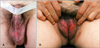
Between 2006 and 2012, 10 Korean patients were treated with our protocol. The characteristics of the 10 patients, 7 men and 3 women, are summarized in Table 1. The patients' ages ranged from 60 to 81 years (mean age: 69.2 years). At the time of treatment, the lesions had been present between 4 months and 30 years (mean: 73.6 months). Clinical manifestations included erythema or eczema with exudation, and crusting and hyperpigmentation of the pubic and genital areas (Fig. 1).
Methods
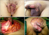
EMPD was diagnosed based on the patients' biopsy results. They had been screened for underlying malignancies and systemic metastases by clinical examinations and appropriate imaging (positron emission tomography-computed tomography, computed tomography etc.). Secondary EMPD was ruled out using immunohistochemistry against markers such as cytokeratin (CK) 7, CK20 and GCDFP15. Multiple scouting biopsies were done with a 3 mm punch preoperatively. The biopsy sites were located 1 cm lateral to the clinically apparent margin. More than 5 biopsy sites were sampled in average, and the results were used to determine the surgical margins. EMPD lesions were excised with surgical margins of 1 cm lateral to the biopsy site when the biopsy result was negative, and 1.5 cm when the biopsy result was positive. The lesions were excised to the depth of the midsubcutaneous fat layer in the pubic area to the dartos muscle in the scrotum and to the superficial fascia of the penis. Histological control by examination of frozen tissue was performed in all the patients. The resultant skin defects were reconstructed with 16/1000 inch split-thickness skin grafts on the penile shaft and mesh grafts on the other sites (Fig. 2). Postoperatively, topical imiquimod was applied every other night, 3 times a week, over the margin and adjacent normal skin for a period of 6 months.
RESULTS
The most frequent location of the EMPD lesions was in the penoscrotal area for male patients and in the vulva for the female patients. None of the patients had underlying malignancies or systemic metastases (Table 1).
The pathologic analysis of the lesions revealed that the epidermis was extensively infiltrated by Paget cells, which are cells found exclusively in the epidermis without dermal invasion, in all the specimens. Immunohistochemical examination of sections using CK7 and CK20 monoclonal antibodies, PAS, D-PAS, and GCDFP15 and Alcian blue PH 2.5, revealed primary EMPD in all the patients (Table 2).
Intraoperative frozen sections were examined to confirm negative margin status.
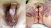
The patients returned for postoperative follow-up examination every 3 months to allow for the evaluation of local or systemic recurrence and genitourinary complications (Fig. 3). All the 10 patients who were followed-up for 1 to 70 months (mean: 34.1 months) remained free of local recurrence and metastases (Table 1).
DISCUSSION
In Korea, EMPD is more common in males than in females (male-female ratio; 3.9:1), which contrasts with findings from studies in Western populations. As a result of this pattern of male predominance, the most common sites of disease are the scrotum, penile shaft, and pubic area5. The patients in the present study also showed a pattern of male predominance and involvement of predilection sites in the penoscrotal area.
Diagnosis of EMPD is confirmed by biopsy. Paget cells are usually identified by their abundant pale-staining cytoplasm and large nuclei, with prominent nucleoli, and typical expression of simple epithelia-type CKs (CK7), sweat gland antigens (EMA, CEA, and GCDFP-15), and mucins (MUC1, and MUC5AC). On the basis of the immunophenotypic heterogeneity of EMPD, 2 distinct types of EMPD have been recognized. Type 1 (endodermal) EMPD cells express an endodermal phenotype (keratin 7+/keratin 20+/GCDFP-15-) and are associated with distant carcinomas. Type 2 (cutaneous and ectodermal) EMPD cells, alternatively, express sweat gland markers (keratin 7+/keratin 20-/GCDFP-15+) and are of cutaneous origin6. According to the immunohistochemical staining results, the patients described in this report showed type 2 cells, leading to the presumption that all of the cases involved primary EMPD. Therefore, extensive examinations for distant carcinomas were not conducted.
There is no consensus on the gold standard treatment for EMPD. Treatment options include topical 5-fluorouracil and topical bleomycin sulfate, radiation therapy, cryotherapy, chemotherapy, CO2 laser therapy and photodynamic therapy7. The histologic extension of EMPD is usually much greater than the clinically apparent margin. Furthermore, EMPD almost exclusively occurs in the anogenital area. Therefore, a paradox confronts surgeons: potentially extensive surgery should be performed in an area where tissue preservation is vital. The most recommended therapeutic mode is complete removal of the lesion by wide excision and complete margin control, using MMS when available. MMS has theoretical advantages for this type of tumor. Lee et al.8 reported that MMS had a recurrence rate of 18.2% versus 36.4% in patients who underwent wide excision. However, compared with surgical excision, the total treatment costs of MMS are significantly higher, mainly owing to the longer surgical time and higher costs of pathological examination9,10. MMS also requires an appropriately equipped procedure room, well-trained nursing staff, and a laboratory with a histotechnician who specializes in MMS tissue processing. In addition, MMS is a highly technical procedure that requires careful attention to detail along with considerable training and experience to achieve the expected high cure rate. Wilson et al.11 reported that MMS incurs higher fees than does wide excision for primary nonmelanoma skin cancer even after adjusting for associated risk factors. In EMPD, treatment cost for MMS is particularly higher than that for wide excision because of the size and complexity of the lesion. Moreover, many EMPD patients are treated by surgeons trained in urology, gynecology, plastic surgery or general surgery without training in MMS. Therefore, the determination of an adequate surgical margin for wide excision is important when MMS is unavailable. Surgical margins are quite variable in the literature because of the rarity of the lesion and the resultant limited experience of authors. Resection margin status is very important because EMPD lesions are irregular and multicentric3. Traditionally, the recommended surgical margin in wide excision is 5 cm12; however, such a large margin may cause serious functional and aesthetic impairment and may not lower the risk of recurrence. Recently, a safe resection margin of 2 cm has been recommended, even though a margin of 1 cm may be sufficient for lesions with clinically clear margins4. In our institution, multiple scouting biopsies 1cm lateral to the clinically evident lesion, have started to be routinely performed before lesional excision13. The purpose of biopsies is to increase preoperative knowledge of the subclinical extent of the tumor. In the present study, the surgical margin was 1 cm lateral to the biopsy site if the biopsy result was negative and 1.5 cm lateral if it was positive. All the specimens were found to be free of tumor cells in the intraoperative frozen specimen examination14.
Imiquimod cream has been thought to be a useful postoperative adjuvant therapy. Imiquimod is a biological response modifier that binds to Toll-like receptor 7 on the cell surface of dendritic cells, macrophages, and monocytes, stimulating both innate and acquired immune function. Some reports about the efficacy of imiquimod in the treatment of EMPD have been recently published15. The aim of postoperative use of EMPD in our study was to improve the chances of cure, reduce patient morbidity, and enhance surgical efficacy. However, considering the high cost of imiquimod and short duration of application, the cost-benefit of imiquimod is not conclusive yet, thus requiring further studies.
The limitation of our study lies in the small number of patients due to the rarity of EMPD in Korea.
We suggest minimal surgical therapy combined with multiple preoperative scouting biopsies and postoperative topical imiquimod as a cost-effective EMPD treatment modality alternative to MMS.


 XML Download