

 PDF
PDF ePub
ePub Citation
 Print
Print


Melanin is produced within melanosomes which later migrate into the melanocyte's dendrite tips using myosin V filaments1 and a dynein "motor"2. In addition, several mechanisms for melanosome transfer from the dendrite tips into the keratinocytes have been suggested, including phagocytosis, release of melanosomes into intercellular spaces followed by endocytosis, direct inoculation ("injection"), and keratinocyte-melanocyte membrane fusion3. Clinically, most of the melanin pigmentary disorders have a strong impact on the quality of life of affected individuals. Especially, many Asian women desire to have flawless skin. Thus, there is a need for the development of skin lightening agents. It is well known that tyrosinase is a rate limiting step in melanin production but melanosome transfer is also a crucial step responsible for melanogenesis. Most hypopigmenting agents are tyrosinase inhibitors and only a few agents are known to inhibit transfer of melanosomes. However, standardized methods to study melanosome transfer are not yet established. Thus, it is necessary to develop a method to study melanosome transfer for the production of hypopigmenting agents.
There are several ways to evaluate melanosome transfer4-6. Confocal fluorescence and electron microscopic examination can be used to study the transfer ability of melanosomes from melanocytes to keratinocytes by labeling melanosme with gold dexrin or melanocytes with green fluorescence dye6. However, these methods are complicated, time consuming, and ineffective for the quantitative analysis of melanosome transfer. Flow cytometry can also be used to study melanosome transfer. In particular, flow cytometry has been applied by double-labeling with fluorescein isothiocyanate (FITC)-conjugated antibody against the melanosomal-associated protein TRP-1, and with Cys5-conjugated antibody against the keratinocyte-specific marker keratin 147. However, these methods were only applied to malignant cells and quantitative analysis was not easy.
In this study, we attempted to develop a simple method to study melanosome transfer by using flow cytometry. Importantly, normal human keratinocytes and normal human melanocytes were used. Then, side scatter (SSC) was analyzed in the cells which showed positivity against the keratinocyte-specific marker keratin 14. SSC is caused by granularity of cells8. If melanosome is transported into keratinocytes, granularity of keratinocytes will be increased. Thus, changes of SSC were analyzed in keratinocytes instead with specific staining against melanosomal proteins. Our methods are easy to apply and not time consuming compared to previous methods. Human keratinocytes were isolated from human foreskins obtained during child circumcision. Skin specimens were processed according to the method of Rheinwald and Green9, as modified in our laboratory using thermolysin (Sigma Chemical Co., St. Louis, MO, USA). Human epidermal melanocytes were also isolated from adolescent foreskins, as previously described10. The cells were maintained in modified MCDB 153 (Sigma) as previously described11, which was supplemented with 5% FBS (Hyclone, Logan, UT, USA), 13 µg/ml bovine pituitary extract (Sigma), 10 ng/ml 12-O-tetradecanoylphorbol-13-acetate (Sigma), 5 µg/ml insulin (Sigma), 0.5 µg/ml transferrin (Sigma), 1 µg/ml tocopherol (Sigma), 0.5 µg/ml hydrocortisone (Sigma), 1 ng/ml human recombinant basic fibroblast growth factor (Sigma), and 1% penicillin-streptomycin (10,000 U/ml and 10,000 µg/ml, respectively) (Gibco BRL, Gaitherbug, MD, USA). The cells were maintained in a humidified incubator with 5% CO2 at 37℃. Third to fifth passage melanocytes were used in the experiments. It is reported that melanocyte-keratinocyte interaction induces calcium signaling and melanin transfer to keratinocytes12. Thus, pigment transfer was decreased when intracellular calcium was inhibited by calcium chelators. To verify the applicability of our method, CaCl2 or Ca2+ chelator were added to the co-culture of keratinocytes and melanocytes.
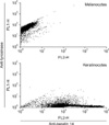
BAPTA-AM (1,2-bis-(o-Aminophenoxy)-ethane-N,N,N'N'-teraacetic acid acetoxymethyl ester, Sigma) is known as a selective chelator of intracellular Ca2+ stores. In order to chelate calcium, cultured keratinocytes were pretreated for 30 min (25 mM of BAPTA-AM) before co-culture of both keratinocytes and melanocytes. In addition, the effects of CaCl2 (2 mM) were also tested. In our study, melanosome transfer was analyzed after 3, 6 and 24 hr incubation. After harvesting the cells, keratinocyes and melanocytes were immunostained with anti-keratin 14 and anti-tyrosinase antibodies. Stained cells were analyzed by flow cytometry. Results showed that cultured keratinocytes and melanocytes well stained with anti-keratin 14 and anti-tyrosinase antibodies, respectively (Fig. 1). When keratinocytes and melanocytes were co-cultured simultaneously, it took 2~3 hr to attach to the culture dishes.
Thus, cells were detached for analysis at 3, 6 and 24 hr after incubation. The antibodies used in this study, were goat polyclonal tyrosinase, 1:100 (sc-7833, Santa Cruz Biotechnology, Santa Cruz, CA, USA); mouse monoclonal keratin 14, 1:100 (MS-115-P1, Thermo Scientific, Fremont, CA, USA); donkey anti-goat IgG-FITC (sc-2024, Santa Cruz Biotechnology); and goat anti-mouse IgG-Phycoerythrin (PE) (sc-3738, Santa Cruz Biotechnology), 1:200. For double staining, antibodies to tyrosinase and keratin 14 were applied together on cells fixed with 4% paraformlaldehyde contained PBS, and cells incubated at 4℃ for 30 min. Next, cells were labeled with FITC- and PE-conjugated secondary antibodies, avoiding direct light at 4℃ for 1 hr. Then, flow cytometric analysis was performed. A total of 10,000 events were collected on the BD FACSCalibur Flow Cytometer (BD Biosciences, San Jose, CA, USA) equipped with CellQuest Pro software for each sample.
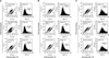
Results showed that there are two groups of cells which showed relatively high anti-keratin 14 and anti-tyrosinase positivity, respectively (Fig. 2). Cells with high anti-keratin 14 were considered to be keratinocytes. Then, these cells were gated (R1) and histogram was analyzed (Fig. 2). In addition, SSC was analyzed. Melanosomes are intracellular organelles. Thus, cells with intracellular organelles can have higher SSC value. Cells were arbitrarily divided into two groups of cells (low SSC: less than 400; high SSC: more than 400). By using 3 hr incubated cells, percentage of high SSC cells was calculated (Fig. 2A, Table 1). Results showed that control cells contain 29.09% high SSC cells. However, in Ca2+ chelator treated cells, high SSC cells represented 22.95%. These results suggest that chelation of Ca2+ may inhibit melanosome transfer in co-culture experimental models. In addition, Ca2+ treated conditions showed a higher percentage (34.99%) of high SSC cells.
Experiments were repeated 3 times and the representative data was shown. These results suggest that short-term incubation of both keratinocytes and melanocytes followed by flow cytometry can be used to study melanosome transfer. Then, experiments were done using cells that were recovered after 6 hr and 24 hr incubation (Fig. 2B, 2C). Data showed that long-term incubation increased percentage of high SSC cells but there were no effects by Ca2+ or Ca2+ chelator (6 hr: control 37.99%, chelated cells 38.81%, Ca2+ treated cells 37.65%; 24 hr: control 76.00%, chelated cells 80.20%, Ca2+ treated cells 80.75%). It showed that long-term incubation increased melanosome transfer but Ca2+ or chelation of Ca2+ did not affect melanosome transfer. Further study is needed to explain why Ca2+ or chelation of Ca2+ can not affect melanosome transfer in long-term incubation (Table 1).
In literature, it is reported that niacinamide gave 35~68% inhibition of melanosome transfer in the co-culture model and reduced cutaneous pigmentation in the skin equivalent model13. However, they used two different keratinocytes and the results were quite various (35%, 68% respectively). In their experiment, they used two different antibodies and analyzed keatinocytes which contain melanosomes. Cells were maintained for 7 days. They also confirmed their results by electron microscopic examination after 3 days incubation. However, co-culture of both keratinocytes and melanocytes may predispose keratinocytes for early differentiation (personal observation). Thus, long-term co-culture of both cells is not easy. Electron microscopic examination is a sensitive way to observe melanosome transfer. However, it is not possible to quantify melanosome transfer. Since previous methods need relatively long-term culture, it will not be easy to have reproducible results. In this study, our methods only need short incubation rather than 3 or 7 day incubation. So, it can keep consistent cell condition especially when toxic agents are treated.
In conclusion, short-term incubation of both normal human keratinocytes and normal human melanocytes was applied to study melanosome transfer by analyzing SSC value. Further study will be required to search for new hypopigmenting agents which inhibit melanosome transfer.

 XML Download