Abstract
A 53-year-old male presented with a 6-year duration of a child's-palm sized hypopigmented patch located on his neck. He had a history of surgical excision of an epidermal cyst on the neck, and the hypopigmented patch developed about one month after the excision next to the surgery site. Application of cold or heat did not make the lesion distinct from the surrounding skin. Pressure on the lesion by a glass slide made the lesion indistinguishable from surrounding uninvolved lesions. Giving friction to the lesion failed to induce erythematous change, making it clearly visible. Histologically, the lesion showed normal findings with adequate numbers of melanocytes in the basal layer. Herein, we present an interesting case of an acquired anemic patch which developed after a cyst excision. We postulate that nerve damage after surgery that regulates the vascular tone of cutaneous vessels may have been an inducing event of the anemic patch in this patient.
Nevus anemicus is a localized cutaneous anomaly that presents as a well-defined hypopigmented patch. It is known to be a congenital non-familial disorder that appears at birth or in early childhood and is usually found on the torso. It is known to be more common in women1,2. We report here a case of an acquired anemic patch that developed in a male in his late forties, after excision of an epidermal cyst.
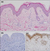
A 53-year-old male presented with a 6-year duration of a hypopigmented patch located on the neck. On physical examination, a child's-palm sized hypopigmented patch was seen on the central aspect of the neck. Wood's light examination showed no accentuation. The patient had a history of surgical excision of an epidermal cyst located on the left lower part of his neck 6 years ago, and the hypopigmented patch developed next to the surgical scar about 1 month after the excision (Fig. 1). Otherwise, he had no medical or familial history. Application of cold or heat did not make the lesion distinct from the surrounding skin. Pressure on the lesion by a glass slide made the lesion indistinguishable from surrounding uninvolved lesions, and giving friction to the lesion failed to induce erythematous change, making it clearly visible from surrounding tissues. The lesion was not transient, and the color of the hypopigmented patch did not show any difference related to postural changes. A punch biopsy was performed, and histologically, the lesion showed normal findings with adequate numbers of melanocytes in the basal layer (Fig. 2a). Staining with melan A and S-100 protein revealed a few melanocytes in the basal layer (Fig. 2b and c).
Nevus anemicus was first described by Vörner3 in 1906, by demonstrating reduced dermographism within the hypopigmented lesion compared to normal skin. It was first thought to be caused by vascular malformation resulting from aplasia of cutaneous vessels, and the importance of vascular or nervous factors in the pathogenesis has been highlighted by Bruner4 in 1912. According to previous pharmacologic studies, nevus anemicus was found to be either due to increased stimulation of the vasoconstrictive fibers or inhibition of the vasodilator fibers of the arterioles. Daniel et al.5 performed a skin graft from lesional skin to normal skin that showed donor dominance with its pale appearance, and it is now thought that nevus anemicus is caused by reduced blood flow due to increased local sensitivity to catecholamines, rather than increased sympathetic stimulation6,7.
Nevus anemicus can be easily diagnosed by demonstrating enhancement of the hypopigmented area upon stroking the involved and adjacent normal skin, which results in erythema only in the adjacent skin, making the lesion clearly visible. Observing an unrecognizable border from the blanched adjacent skin by a diascopic examination can also help in diagnosing nevus anemicus. Nevus anemicus can clinically resemble various hypopigmented disorders such as vitiligo, nevus depigmentosus, and Bier's spots. Vitiligo and nevus depigmentosus can be excluded on the basis of clinical and histological features. Wood's light examination, and stroking the skin can be useful as diagnostic techniques for differential diagnosis. Histologically, nevus anemicus shows normal findings and can be distinguished from the findings of vitiligo, with decreased numbers or absence of melanocytes, and nevus depigmentosus, with a normal number of melanocytes but abnormal melanization. Bier's spots, which are transient anemic macules, are considered to be an exaggerated physiological response of cutaneous small vessels to venous hypertension8. It is usually seen on the extremities, and if the venous stasis is reduced by raising extremities or taking off the tourniquet, the spots tend to disappear9.
The anemic patch that had developed in this patient appeared on the central aspect of the neck 1 month after a surgery. He did not have any history of a hypopigmented lesion on the neck before the surgery, and therefore, it can be defined as an acquired rather than a congenital lesion. Furthermore, the hypopigmented patch in this patient was considered to occur as a vessel origin and not by melanocyte origin, considering that wood's light examination failed to accentuate the lesion. Also,when pressure was given by a glass slide, the lesion became indistinguishable from the surrounding uninvolved lesions. Observation of melanocytes in the basal layer after melanocyte staining also supports the fact that the anemic patch in this patient developed by vascular origin. Thus, although the exact mechanism of the pathophysiology is unknown, the development of the acquired anemic patch can be postulated to have been derived from the surgical procedure, which may have been an inducing event. As the lesion developed next to the surgical scar and not on the surgery site itself, it can be assumed that the lesion was not simply caused by decreased blood flow according to vascular damage on the surgery site, but due to the effect of nerve damage during the surgery that regulates the vascular tone of cutaneous vessels.
Although several types of acquired hypopigmentated disorders can appear in adults10, there have not been any previous cases of acquired anemic patches related to an excision reported in the literature. Despite the existing controversy of the diagnosis, the acquired anemic patch developed in our patient showed several physical and histologic characteristics similar to nevus anemicus. Therefore, the acquired anemic patch that developed after a cyst excision in this case can be explained as a variant of nevus anemicus.
Figures and Tables
References
2. Miura Y, Tajima S, Ishibashi A, Hata Y. Multiple anemic macules on the arms: a variant form of nevus anemicus? Dermatology. 2000. 201:180–183.

4. Bruner E. Naevus anaemicus. Gaz Lek. 1912. 32:363–368.
5. Daniel RH, Hubler WR, Wolf JE, Holder WR. Nevus anemicus. Donor-dominant defect. Arch Dermatol. 1977. 113:53–56.

7. Mountcastle EA, Diestelmeier MR, Lupton GP. Nevus anemicus. J Am Acad Dermatol. 1986. 14:628–632.


 PDF
PDF ePub
ePub Citation
 Print
Print


 XML Download