

 PDF
PDF ePub
ePub Citation
 Print
Print


INTRODUCTION
Skin contributes to the first line of defense against any foreign materials outside of our body, as a physical barrier. As immune sentinels, keratinocytes can recognize foreign or danger stimuli from the outside via pattern-recognition receptors, such as Toll-like receptors (TLRs), and release innate immune mediators, including cytokines and chemokines, under the stimulation of the keratinocytes. Epidermal keratinocytes express several TLRs, including TLR1, TLR2, TLR3, TLR4, TLR5, TLR6, TLR7 and TLR91, and through the TLRs, keratinocytes initiate and promote immune responses in the skin.
For inflammatory skin disease, pathogenesis is due to the sum of the immune reactions of the keratinocytes, immune cells and soluble mediators. The three main types of CD4+ T cells can be found in the skin during inflammatory skin disease, i.e., Th1, Th2 and Th17. For example, Th1-dominant immune reactions were reported to be associated with the autoimmunity or psoriasis, and Th2-dominant responses were related to that of asthma or atopic dermatitis. The Th17 cells were reported to contribute to defend against various fungal or bacterial infections, and possibly induced atopic dermatitis and epidermal changes in psoriasis2,3 via secretion of interleukin (IL)-17 and IL-22. IL-17 and IL-22 increased antimicrobial peptides, β-defensins and cathelicidins, from keratinocytes4. In addition to the antimicrobial peptides, keratinocytes secrete cytokines, including IL-1, IL-6, IL-10, IL-18 and tumor necrosis factor (TNF)5.
Spa therapy is widely used for the treatment of inflammatory skin diseases, such as atopic dermatitis, psoriasis, pruritus, rosacea, seborrheic dermatitis and others6. The efficacy of spa therapy for inflammatory skin diseases and the mechanisms are only partly understood, and presumably incorporate chemical, thermal, mechanical and immunomodulatory effects7,8. Among them, we investigated the immunomodulatory or anti-inflammatory effect of thermal spring water on the expression of pro-inflammatory cytokines in the HaCaT cells under TLR stimulation, as well as the effect on differentiation of CD4+ T cells under spring water.
MATERIALS AND METHODS
Cell culture
HaCaT (human keratinocyte cell line) was kindly provided by Dr. Tae-Yoon Kim (College of Medicine, The Catholic University of Korea) and was cultured in Dulbeco's Modified Eagle Medium (DMEM, Gibco-BRL, Grand Island, NY, USA) supplemented with 10% fetal bovine serum (FBS, Gibco- BRL) and 100 U/ml penicillin/streptomycin (Gibco- BRL), at 37℃ in an incubator containing 5% CO2.
Preparation of spa spring water
Water was collected from the Yong-gung oncheon (Incheon-si, Gangwha-gun, Korea) spring and filtered through 0.44 um filters. Waters were stored at 4℃, until used for the experiments. For the measurement of water osmolarity, we used a Micro-Osmometer 210 (FISKE Associate, Norwood, MA, USA).
MTT assay
For the 3-(4,5-dimethylthizol-2-yl) 2,5-diphenyl tetrazolium bromide (MTT, Sigma-Aldrich, St. Louis, MO, USA) assay, cells were plated onto 96-well microtiter plates at a density of 3×10⁴/200 µl in fresh medium, and then treated with hot spring water at a serially diluted concentration. Cells were cultured for 1, 4, 10, and 24 hours, respectively, to observe a time dependent effect. After the indicated time, 20 µl of MTT (5 mg/ml in phosphate-buffered saline) was added to each well, and the plates were returned to the incubator for an additional 4 hours. At the end of the incubation period, the supernatants were discarded by a suction and 200 µl dimethyl sulfoxide was added to all the wells, in order to dissolve the dark blue formazan crystals. The plates were subsequently covered with aluminum foil, gently shaken for 15 minutes, and read at a wavelength of 570 nm.
TLR stimulation
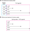
TLR agonists were treated with the following final concentrations for 24 hours. Tripamitoyl-S-glyceryl-cysteine (Pam3Cys, 1 µl/ml), heat-killed Listeria monocytogenes (HKLM, 106 cells/ml), polyriboinosinic polyribocytidylic acid (poly (I : C), 10 µl/ml), lipopolysaccharide (LPS, 10 µl/ml), flagellin (10 µl/ml), and Pam2CGDPKHPKSF (FSL-1, 1 µl/m) were from InvivoGen (San Diego, CA, USA). Spa spring waters were added simultaneously or pre-treated 2 hours before the TLR agonist treatment. Cells were cultured for 1, 4, 10, or 24 hours in each treatment group.
ELISA
Levels of IL-6, IL-8, granulocyte-macrophage colony-stimulating factor (GM-CSF), and TNF-α (BD OptEIA™, BD Biosciences Pharmingen, San Diego, CA, USA), IL-1α (Biolegend, San Diego, CA, USA) in HaCaT supernatant treated with TLR agonist in the presence or absence of hot spring water were quantified according to the manufacturer's protocol. IL-6 and TNF-α in mouse antigen presenting cells (APC) supernatant were also measured. In brief, the wells were coated with 100 µl of capture antibody in coating buffer (0.1 M sodium carbonate, pH 9.5) and the plates were incubated overnight at 4℃. After washing the wells with washing buffer (phosphate buffered saline [PBS] with 0.05% Tween-20), the wells were blocked with assay diluents (10% FBS in PBS) for 1 hour at room temperature (RT), followed by the addition of 100 µl/well of cell supernatant and cytokine standard solutions for 2 hours at RT. After washing, 100 µl of detection antibody and streptavidin-conjugated horseradish peroxidase reagent were added to the wells, which was then incubated for 1 hour at RT. After extensive washing, 100 µl of substrate solution (tetramethylbenzidine and hydrogen peroxide, BD Biosciences Pharmingen) was added to each well and the plates were incubated for 30 minutes at RT, in darkness. Stop solutions (2 N H2SO4) were added, and the absorbance was read at 450 nm within 30 minutes.
Mouse spleen cell isolation
Naïve CD4+ T cells were purified from the mouse spleens, via magnetic isolation (Miltenyi Biotec GmbH, Gladbach, Germany). For the preparation of the spleen cell suspensions, spleens from 8 weeks-old female Balb/c mice were removed and minced with a Nylon mesh (70 µm pore). After the cells were pelleted, erythrocytes were lysed using hypotonic buffer (0.15 M NH4Cl, 10 mM KHCO3, 0.1 mM Na2EDTA). The cells were washed in PBS and incubated with anti-CD4 antibody for 15 minutes, at 4℃. Directly following, the cells were conducted onto a magnetic separator to isolate the CD4+ cells and collected with positive selection. CD4- cells were seeded onto a culture dish of 100 mm in diameter and incubated for 4 hours for the collection of the adherent cells (used as APC in these experiments) by gentle pipetting.
CD4+ T cell differentiation
CD4+ naïve cells were seeded at a density of 2×10⁵ per well in 96-well plates, and were cultured in DMEM, containing 10% FBS. For each of the helper T cell differentiation, the skewing conditions are as followed; IL-12 (25 ng/ml, Biolegend) and anti-CD28 (1 µg/ml, Biolegend) for Th1, IL-4 (25 ng/ml, Biolegend) and anti-CD28 (1 µg/ml, Biolegend) for Th2, IL-6 (25 ng/ml, Biolegend), transforming growth factor (TGF)-β (2 ng/ml, R&D system) and anti-CD28 (1 µg/ml, Biolegend) for Th17, IL-2 (25 ng/ml, Biolegend) and anti-CD28 (1 µg/ml, Biolegend) for T reg. All cells stimulated with anti-CD3 antibodies with serial dilutions, in which the initial concentration was 3 µg/ml. Cells were incubated for 3 days in the presence or absence of spa spring water.
Cell proliferation assay
Before seeding for the differentiation of each of the helper T cell differentiation, the CD4+ cells were labeled with carboxyfluorescein diacetate, succinimidyl ester (CFSE, CellTrace™ CFSE Cell Prolifetration kit, Invitrogen, Paisley, UK). First, the cells were resuspended in prewarmed PBS/0.1% bovine serum albumin, at a concentration of 106 cells/ml, and 10 µM of CFSE were added. After incubation at 37℃ for 10 minutes, 5 volumes of ice-cold culture media was added to the cells to quench the staining, incubated for 5 minutes on ice, and pelleted by centrifugation. The cells were resuspended in fresh media for a total of three washes, and seeded under each condition for the differentiation of Th1, Th2, Th17, and Treg cells. After 3 days, CD4+ and CFSE+ cells were measured for the degree of proliferation, via a flow cytometry, and were analyzed using ModFit LT software (Verity Software House, Topsham, ME, USA), based on the reduction of CFSE positive cells.
Flow cytometry
APCs were stimulated with TLR3 agonist, poly (I : C) (10 µg/ml) for 24 hours with or without spa spring waters. APCs stimulated with poly (I : C) with or without hot spring water were collected, washed and stained with anti-mouse I-A/I-E (Biolegend, clone M5/114.15.2, Rat IgG2b, κ) to analyze the expression of MHC II on the surface of APCs for 20 minutes, at RT. Samples were acquired on FACSCalibur system (BD Bioscience, San Jose, CA, USA) and were analyzed using CellQuest software (BD Bioscience).
RESULTS
Determination of dilution concentration of spa spring water for HaCaT cell culture
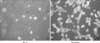
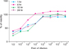
Because the osmolarity of spa spring water from Yong-gung oncheon was 700 mOsm/kg, the HaCaT cells were not able to maintain the growth in undiluted culture medium (Fig. 1). Therefore, we first determined to set an optimal dilution concentration for HaCaT culture conditions by the MTT assay. For this, HaCaT cells were cultured with spa spring water at a concentration of serially 2-fold dilution for 1, 4, 10, and 24 hours and the minimum concentration of spa spring water was determined. We found that each group of cells was up to 100% of viability under a 1 : 32 fold dilution (Fig. 2).
Cytokine production of HaCaT cells under the TLR agonist treatment with spa spring water
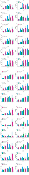
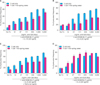
TLRs act as primary sensors that detect a wide variety of microbial components and elicit innate immune response, which guides the acquired immunity. Therefore, keratinocyte constitutively expressing TLRs may affect skin immune response, by regulating the inflammatory responses, via TLR signaling. We attempted to determine whether the spa spring water affected the TLR stimulated production of pro-inflammatory cytokines, including IL-1α, IL-6, IL-8, TNF-α, and GM-CSF, on the HaCaT cells. TLR1 agonist, through TLR6 agonist treatment, induced the attenuation of cytokine production in the exposure to spa spring in advance or simultaneously with TLR agonist treatment (Fig. 3, 4).
Proliferation characteristics of T cells under spa spring water
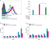
We subsequently attempted to ascertain the potential involvement of spa spring water in the differentiation of the helper T cells. Because the effector T cells, such as Th1, Th2, and Th17 cells act on various skin resident cells and potentiate the immune reaction, regulating T cell differentiation may modulate pro-inflammatory responses. To evaluate the in vitro suppressive capacity of spa spring water toward the helper T cell differentiation, magnetic sorted CD4+ T cells, from the spleen, were prepared from Balb/c mice. CFSE-labeled CD4+ T cells were cultured under various conditions, which polarized Th1, Th2, Th17, and Treg with or without spa spring water. After 3 days, the cells were harvested and stained with CD4 and interferon (IFN)-γ for Th1, IL-4 for Th2, IL-17 for Th17 and Foxp3 for Treg cells, respectively. Then, the flow cytometry was employed to evaluate the precursor frequency in terms of the T cell differentiation. As shown in Fig. 5, CD4+ naïve T cells were profoundly proliferated and differentiated under each skewing condition, along with anti-CD3 stimulation. Spa spring water suppressed the proliferation of Th1, Th2 and Th17 cells. In contrast to the suppressive effect on Th1, Th2 and Th17 cells, proliferation and differentiation to Treg cells were promoted under spa spring water treatment. These results indicate that spa spring water may affect the distribution of the helper T cells in the immune response, by suppressing the polarization of the Th1, Th2 or Th17 cells.
Cytokine production and major histocompatibility complex (MHC) II molecule expression of APCs under TLR3 agonist treatment were affected by spa spring water
Skin immunity is achieved by an interaction of different kinds of immune cells. In particular, APCs, such as Langerhans cells found in the epidermis, are the best-characterized dendritic cell population. They have the ability to process antigens in the periphery, transport it to the draining lymph nodes, where they are able to cluster with, and activate the antigen-specific naive T cells. APCs in the dermis may also provide alternative routes of antigen presentation, which can be important in the regulation of skin immune responses. Therefore, APCs are vital for the induction of immune responses to antigens encountered via the skin. To determine whether the expression of MHC II on the surface of APC is modulated by spa spring water, we isolated APC from BALB/C mouse spleen, and stimulated it with 20 µg/ml of poly (I : C) for 24 hours. TLR3 stimulation via poly (I : C) strongly induced MHC II expression on the APCs. By contrasts, APC treated spa spring water down-regulated the surface level of class II MHC expression, under TLR 3 stimulation (Fig. 6A). In addition, we observed that TLR3 agonist, poly (I : C) is a very potent inducer of inflammatory responses in the HaCaT cells9. Therefore, we measured and compared poly (I : C) mediated production of inflammatory cytokines from the APCs with or without spa spring water (Fig. 6B, C). APC induced TNF-α and IL-6 under poly (I : C) stimulation and cytokine production was reduced in the presence of spa spring water. These results showed that TLR-triggered inflammatory responses in APCs might also be modulated under spa spring water treatment.
DISCUSSION
In this study, we evaluated the effect of spa spring water from Yong-gung oncheon on cells related to the skin immune reaction. The treatment of spa spring water from Yong-gung oncheon decreased the expression of proinflammatory cytokines under TLR stimulation to the HaCaT cells and APCs. In addition, spa spring water attenuated the differentiation process of subsets of T helper cells, i.e., Th1, Th2 and Th17 cells.
The therapeutic mechanism of spa spring water in dermatologic diseases can be divided into three categories: active ingredient, thermal effects and mechanical effects. Minerals in spa spring water, such as sulfur, magnesium, calcium or selenium induce anti-inflammatory, keratolytic, antibacterial or antifungal effects6. For anti-inflammatory effects, the inhibition of Th1 differentiation, inhibition of cytokine production from keratinocytes, and modulatory effects on epidermal Langerhans cells have been reported10. In this study, we observed the significant inhibitory effect on the secretion of pro-inflammatory cytokines, including IL-6, IL-8, IL-1α, TNFα and GM-CSF from keratinocytes (Fig. 4). In addition, we observed the differentiation of Th1, Th2, or Th17, but not of Foxp3+ Treg cells under the treatment with water from Yong-gung oncheon (Fig. 5).
Th17 cells are related to the autoimmunity or inflammatory skin disease, such as psoriasis or atopic dermatitis via IL-17 and IL-22 secretion. IL-17 acts directly on keratinocytes and induces the production of MIP-3α, IL-8 and β-defensin, whereas, IL-22 regulates the keratinocyte differentiation11,12. In addition, IL-17 promotes the neutrophil recruitment by inducing the neutrophil-attracting chemokines (CXCL1, CXCL2, CXCL5, and CXCL8) and stimulates neutrophil production by inducing granulopoiesis factors (G-CSF or GM-CSF)13. In case of Staphylococcus aureus infection, the role of Th17 was reported to be critical: in mice bacterial clearance was impaired in IL-17R-deficient mice, and in human hyper immunoglobulin E syndrome, atopic dermatitis, human immunodeficiency virus/acquired immune deficiency syndrome or mucocutaneous candidiasis, in which there is a common deficiency of Th17 cells. These IL-17-mediated defenses against S. aureus infection are involved with promotion of neutrophil recruitment via cytokines, chemokines or adhesion molecules and production of antimicrobial peptides. In this context, reduced Th17 cell differentiation, by spa spring water, implies that spa spring water may reduce the immune reaction in the epidermal layer, partly by affecting the antimicrobial peptide production and epidermal differentiation irrespective of antibacterial effect of IL-17.
Regulatory T cells (Treg cells) mediate immunosuppression and tolerogenic responses through contact-dependent or -independent mechanisms14-16. Foxp3+ Tregs produce IL-10 or TGF-β as effector molecules, and the balance between Treg cells and effector T cells is crucial for the maintenance of homeostasis and self-tolerance17. In this study, spa spring water from Yong-gung oncheon treatment induced Foxp3+ Treg cell differentiation in vitro, implying that the immune modulatory effect of spa spring water also includes Treg cell-induced immune suppressive effects.
TLRs are a type of pattern recognition receptor, which recognize microbial products known as pathogen-associated molecular patterns, i.e., bacterial lipoproteins, zymosan, LPS, flagellin, ssRNA, dsRNA, and unmethylated CpG DNA. TLRs are transmembrane receptors and present on the cell surface or on the surface of endosomal compartments. It was reported that TLR3 ligand poly (I : C) was the most potent stimulator of IL-8, IL-6 and TNFα secretion18 in the primary keratinocyte and HaCaT cell lines. In this experiment, we used TLR1 to TLR6 agonists to activate the HaCaT cells inducing pro-inflammatory cytokines. We observed that spa spring water treatment led to reduced IL-6, IL-8, TNF-α, IL-1α, and GM-CSF from the HaCaT cells, following TLR ligand treatments, and spa spring water from Yong-gung oncheon suppressed the expression of IL-6, TNF-α and class II MHC expression of APC in this experiment (Fig. 6). In case of atopic asthma, IL-5, IL-13, IL-1β, IFN-γ, IL-12, GM-CSF, IL-4, and IL-10 were elevated, compared with those of the non-atopic patients19. Further studies are required to compare the cytokine profile in non-atopic and atopic dermatitis patients after a spa treatment.
These results showed that spa spring water treatment suppressed the inflammatory cytokines production, and also modulated differentiation of CD4+ T cells into Th1, Th2, and Th17 cells, but not Tregs cells. With these experimental protocols, we can evaluate and compare the efficacy of spa spring waters in Korea, immunologically. Furthermore, we would define immune-active ingredient from this spa spring water to reveal immune modulatory mechanism of spa spring waters in Korea.
 XML Download