

 PDF
PDF ePub
ePub Citation
 Print
Print


INTRODUCTION
Acne vulgaris is a common skin disease, affecting more than 85% of adolescents, often continuing into adulthood1,2. Currently, while oral and topical antibiotics and retinoids represent the most conventional, widely-accepted pharmacologic therapies for acne, both have significant side effects: widespread use of antibiotics increases the risk of resistant bacterial strains, while oral isotretinoin has been linked to dry skin, headaches, fetal defects and depression. Alternatives to pharmacologic therapies include chemical and physical exfoliation techniques and light devices, most notably blue light, intense pulsed light (IPL), light-emitting diodes, various lasers (especially infrared) and photodynamic therapy (PDT). Interestingly, while IPL with or without PDT has proven effective for treating acne in patients of European descent, no significant improvements were observed in studies among Asians. Kawana et al.3 propose that this discrepancy results from the use of inappropriate wavelengths or inaccurate irradiating light source targeting. These authors also show that IPL using dominant wavelengths of 400 nm to 700 nm was effective in reducing acne vulgaris lesions among Asians. However, treatment with IPL is often poorly tolerated, with many subjects reporting pain (associated both with the IPL treatment and the topical anesthesia), immediate erythema, and sensations of burning and/or stinging. Additionally, rare episodes of crusting, bulla formation and hyperpigmentation have been reported after IPL treatment.
Recently, a novel device (Isolaz, Aesthera Co., Pleasanton, CA, USA) that combines vacuum pressure with a broadband light source (400 nm to 1,200 nm) was developed for the treatment of acne. Unlike other devices that are currently available, this device uses gentle pneumatic energy to draw the target tissue into the treatment tip, with negative pressure lifting the sebaceous gland and thus bringing it closer to the surface of the skin4. The vacuum then elevates and everts the sebaceous gland, allowing it to open up and empty its contents, ejecting the acne-causing bacteria, sebum, dead skin cells, and other impurities onto the surface of the skin. Such photopneumatic devices are the only lasers approved by the United States Food and Drug Administration for the treatment of comedonal and pustular acne, as well as inflammatory acne. The purpose of this study was to determine the clinical efficacy and safety of photopneumatic therapy for the treatment of acne vulgaris of the face in an Asian sample.
MATERIALS AND METHODS
Patients
All components of this study were performed at the Kyung Hee University School of Medicine, Department of Dermatology, located in Seoul, Korea. The protocol adhered to the Helsinki guidelines, and the study underwent review and approval by the Kyung Hee University Institutional Review Board (KHUHMDIRB1105-01). Informed consent was obtained from all subjects prior to any study-related procedures. In total, 20 Korean patients with inflammatory acne vulgaris of the face were subjected to photopneumatic therapy between July 2010 and January 2011. For this study, exclusion criteria included concurrent pregnancy or lactation, the use of any photosensitizing drugs, a prior history of porphyria or photosensitivity, or oral antibiotic therapy at any point during the course of study. All topical and oral acne medications were discontinued 3 months prior to study enrollment, and no oral or topical acne medications were permitted during the study.
Study design and laser treatment
In this split-face controlled study, 10 patients received treatment on the right side of the face and 10 on the left side of the face, with the untreated side of each subject's face serving as a control. The subject's facial skin was first cleaned with a mild soap and water in order to remove any cosmetics or debris. A portable photopneumatic device (Isolaz) was used for all treatment sessions. No topical or systemic anesthetics were administered. For most treatments, the power was set at 6 (approximately equivalent to 5.2 J/cm2) and the vacuum at iMP (delivers multiple vacuum pulses in each cycle). A large tip (15×30 mm) was employed for treatments of the cheeks and a small tip (5×12 mm) for the nose. Typically, patients were treated with 1 pass during each of the four treatment sessions that occurred at 2 week intervals. Patients were assessed at three follow up visits 4, 8 and 12 weeks after the final treatment session.
Evaluation
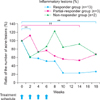
Prior to each treatment session and during each follow-up visit, two different investigators manually counted the number of acne lesions for each patient on both the treatment and control sites. Clinical photographs were obtained at each of these time-points for evaluation purposes. Other clinical observations, including erythema, purpura, and treatment-associated pain level, were recorded by research staff. After each treatment session, all skin lesions were compared to pretreatment appearance and lesions on the contralateral side. Using these data, patients were then divided into three groups based on the clinical improvement seen in inflammatory acne lesions: responders, partial responders, and nonresponders. To quantify the actual degree of improvement, the ratio of remain acne lesions at each treatment to the initial acne lesion was calculated after the first, second, and third treatments. Responders were defined as showing a reduction in the ratio of inflammatory acne equal to or greater than 50%, partial responders as showing a reduction less than 50%, and nonresponders as showing a reduction less than 25%.
Wood's light examination
All patients were examined using a Wood's light both before and after treatment. Photos were also obtained under these conditions before each treatment session, so that the levels of porphyrin fluorescence could be compared to the subsequent set of photos by the clinic staff.
Patient self assessment
At 12 weeks after the final treatment session, patients assessed the improvement in their acne as one of the following: 'marked improvement,' 'moderate improvement,' 'slight improvement,' 'no change,' or 'worse.' Additionally, patients were asked whether they would recommend this particular treatment modality to others.
RESULTS
Patients
Although 20 subjects were initially enrolled in the study, 2 dropped out prior to study completion. One dropped out after the second treatment session due to a scheduling conflict, while the other refused further treatments after the third session, stating that the treatment was not effective. The remaining 18 subjects completed the entire study treatment protocol and all follow-up visits. Patients ranged in age from 24 years to 34 years, with an average age of 27.5 years, with 18 females and 2 males. Fitzpatrick skin types 3 through 5 were represented in our cohort. According to Korean acne grading system, there were 5 patients with grade 1, 12 patients with grade 2 and 3 patients with grade 3. Prior to enrollment, all patients had previously failed topical and systemic treatment or received suboptimal benefit from these agents. After the final photopneumatic treatment session, 9 patients requested additional photopneumatic treatments for the contralateral side of the face, which were subsequently performed at 2 week intervals. The remaining patients did not receive any further photopneumatic treatment on either side of the face.
Physician evaluated acne counts
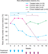
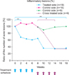
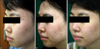
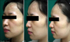
Almost all of the patients experienced a reduction both in inflammatory and noninflammatory lesion counts on the treated sides of the face: the mean inflammatory papule and noninflammatory papule counts were significantly lower on the treated side than the untreated side. The treatment-associated pain was well tolerated even without topical anesthesia. On the treated side, the number of noninflammatory acne lesions decreased to 43.83% of the pretreatment value after the fourth treatment (p<0.01) and to 41.38% at the final follow-up visit (p<0.01) (Fig. 1). Similarly, the inflammatory lesion count decreased to 64.7% of the pretreatment value on the treated side after the fourth treatment (p<0.01), and to 37.5% by the study end (p<0.01) (Fig. 2). When stratified by therapy response grouping, 13 patients fit the criteria for the responder group, 3 patients for the partial responder group, and 2 patients for the nonresponder group (Fig. 3, 4). Among the responder group, the number of inflammatory acne lesions decreased to 23.5% of the pretreatment value at the 10 week follow-up visit (p<0.01) and to 26.5% at the final follow-up visit (p<0.01) (Fig. 5). In patients who underwent additional treatment on the contralateral face, a marked improvement was observed in both inflammatory and noninflammatory lesions, significantly different from untreated side of face (p<0.05). Two patients reported mild pain during treatment and one experienced immediate erythema. Two additional subjects complained of an exacerbation of their acne, while another developed post-treatment petechiae. Crusting, bulla formation, or hyperpigmentation were not observed among any enrolled subjects.
Wood's light examination
On examination with the Wood's light, no immediate difference in red fluorescence was detected between the pre- and post-treatment photos. When later compared to photos obtained during follow-up visits, some patients did show decreased levels of red fluorescence; however almost all were equivocal. Initial extent and density of red fluorescence showed no relation to degree of improvement of acne after treatment.
Patient assessment
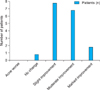
The data for the patient-assessed treatment outcomes at the 12 week follow-up visit are summarized in Fig. 6. Regarding the acne on the treated side of the face, a 'marked improvement' was reported by 2 patients, a 'moderate improvement' by 7 patients, a 'slight improvement' by 8 patients, and 'no change' by 1 patient (Fig. 6). Furthermore, 16 patients reported that they were willing to recommend this treatment to others, while 2 stated that they would not recommend photopneumatic therapy due to its low perceived efficacy.
DISCUSSION
Significant improvements were observed in acne lesions on the treated sides of patients' faces and a significant reduction in the total number of acne lesions was observed on the treated side when compared with the untreated side. These data also indicate that after the final treatment session, measurable improvements were noted in inflammatory lesions on the treated side of the face, an effect that persisted to the final follow-up visit at 12 weeks.
Among the individuals who opted for additional treatment on the contralateral side of the face, a marked improvement was seen in both inflammatory and non-inflammatory acne lesions, a difference that reached statistical significance when compared with the state before treatment. We contend that the greater degree of improvement observed among these individuals results primarily from self selection: subjects who achieved effective treatment results were more inclined to request additional treatments on the untreated side.
No severe side effects occurred during this study. In fact, the only adverse events reported were transient erythema, purpura and acne flare. The petechiae that occurred in one patient developed in an area between the nasal bridge and inner canthus. Although all evidence of petechiae had vanished within 1 week after treatment, we did not subject this area to treatment in any other patients, and would advise other providers to use caution when treating this anatomic region. According to Wanitphakdeedecha et al.5, most patients experienced acne worsening early in the treatment course during their photopneumatic treatment study. They suggest that causes of lesional worsening include incomplete comdone removal or comedonal rupture during application of negative pressure, leading to development of tissue inflammation and inflammatory lesions. Five patients showed acne flare after 1st treatment session at our study (Fig. 4).
Interestingly, while several articles have suggested that photopneumatic therapy is an effective method for evacuating sebum, we rarely observed expelled sebum at the cheek area that is main site of acne. The expelled sebums were occasionally observed at the nose area where there was a pre-existing dilated pore. Nonetheless, significant reductions in noninflammatory acne were observed after photopneumatic treatment despite the lack of any obvious sebum extraction. We contend that the therapeutic mechanism of photopneumatic device on acne lesions is more closely related to the phototherapeutic effect of the device rather than the pure physical mechanics.
Several studies3 have suggested that IPL is not an effective treatment modality for acne in patients of Asian descent. However, these studies employed IPL device light sources with wavelengths set at greater than 560 nm. Furthermore, such devices only emitted light in the yellow and red regions (550~800 nm), thus lacking any blue region wavelengths (400~500 nm). Conversely, the study by Kawana et al.3 demonstrated that IPL (400~700 nm and 870~1,200 nm, 13 J/cm2, 5 sessions at 1 week interval) does have a potent therapeutic effect on both inflammatory and non-inflammatory acne lesions in Asians. Our device has a broadband light source (400 nm to 1,200 nm), thereby including all blue light regions6.
Coproporphyrin III, the major endogenous porphyrin of Propionibacterium acnes, absorbs light in both the ultraviolet A (320~400 nm) and blue light spectra, with a maximum absorption near 415 nm7. Accordingly, different ranges of light can produce vastly variable results. Although porphyrin absorption is strongest in the blue spectrum, these wavelengths penetrate the dermis poorly in vivo. Facial sebaceous glands, one component of the follicular unit, are located approximately 0.5 mm to 1.0 mm below the cutaneous surface, thus the vacuum suction delivered by the photopneumatic device allows deeper light penetration than would be obtained with traditional blue light therapy. That wavelengths in the red light spectrum are significantly longer which may explain the greater efficacy of the IPL device. Similarly, there is ultrastructural evidence for thermal damage to pilosebaceous units and bacteria by the photopneumatic device8. Mechanical extrusion of comedonal contents from the infundibulum has been observed histologically. Although we rarely found notable sebum removal by the vacuum mechanism at cheek area, it may have facilitated deeper light penetration in addition to minimal mechanical effect, thus reducing side effects and increasing treatment effectiveness. The combination of thermal effect, mechanical effect and vacuum effect might act comprehensively on the acne lesion.
Gold and Biron9 describe a study of seven patients with mild to moderate acne vulgaris who received 4 treatments with photopneumatic therapy at 3 week intervals. By the study end, significant reductions in both inflammatory and noninflammatory lesions had occurred at 3 months. In another study by Wanitphakdeedecha et al.5 photopneumatic therapy was administered to 20 patients at 2 week intervals, after which time a modest reduction in acne lesion counts and global clinical improvement were observed in the majority of patients. In both articles, no severe side effects occurred, with the most common adverse event encountered being mild pain and transient erythema.
However, no split-face trials were previously published. Therefore, by using a split-face format, we can definitively determine that any observed improvements in acne are the direct result of the photopneumatic treatment. We contend that photopneumatic treatment is an effective alternative treatment method for acne patients who must avoid oral medications. Busy patients may also especially benefit from this device because it can be used without topical anesthesia, and therefore significantly less time is required than with many other laser-based protocols. Compared to IPL therapy, there is virtually no blistering, burning or postinflammatory hyperpigmentation associated with photopneumatic therapy. Photopneumatic therapy is significantly more safe and effective. One limitation of this study is the small number patients enrolled, and additional; larger trials are clearly needed.
 XML Download