

 PDF
PDF ePub
ePub Citation
 Print
Print


INTRODUCTION
Scabies is a contagious skin infestation caused by the penetration of the obligate human parasitic mite Sacrcoptes scabiei var hominis into the epidermis. Animal scabies such as canine sarcoptic mange or feline mange may afflict humans with a pruritic dermatosis with excoriation and crusting. However, it is considered to be rare because of the relative host specificity of the mites1,2.
Human scabies is characterized clinically by pruritus with nocturnal exacerbation and contagiousness. Also, scabietic nodules as well as visible skin burrows can be the pathognomic lesions of scabies2. Under normal circumstances, the diagnosis can be readily made by the clinical distribution, appearance of skin lesions and clinical history. However, scabies can present with an atypical clinical pattern in some certain situations, including patients on prolonged steroid therapy, immunocompromised condition and nodular scabies. Also, diagnosing scabies in infants or the elderly can be challenging since the clinical features in these age groups may differ from those of normal adults3. Given that most dermatologists rely on the clinical findings or skin scraping in diagnosing scabies, it can be difficult to distinguish scabies from other dermatoses in such cases.
Dermoscopy was originally described by dermatologists as a useful tool for the different diagnosis of the pigmented lesions and melanoma. Dermoscopy was also viewed as a sensitive and handy tool in diagnosing scabies in vivo4. It permits the identification of a triangular or V-shaped structure corresponding to the fore portion of the mite, including the head and the the pairs of legs. Since the mite is usually found at the end of the burrow, it has been described as resembling a jetliner with its trail, a delta glider, or a spermatozoid2,5.
There have been many case reports about the experiences of diagnosing scabies with dermoscopy. The weight of this evidence is that dermoscopy is very useful, especially in certain circumstances. Walter et al.6 and Dupuy et al.7 evaluated the diagnostic characteristic of dermoscopy. However, to our knowledge, there have been few well-designed clinical studies concerning the diagnostic accuracy of dermoscopy by comparing skin scraping "with dermoscopy" to skin scraping "without it".
To address this shortcoming, we designed this prospective, evaluator-blinded study, in which the diagnostic accuracy of dermoscopy for diagnosing scabies was evaluated, and demonstrated the specific circumstances in which dermoscopic identification ("with dermoscopy") is more useful in diagnosing scabies. Lastly, we identified the specific clinical features that can be used as one of the positive markers in diagnosing scabies.
MATERIALS AND METHODS
Patients
From January 1 to December 31, 2010, patients in the Department of Dermatology of Kangdong Sacred Heart Hospital were eligible to have their clinical data recorded if there was a clinical suspicion of scabies (presence of characteristic symptoms of scabies including pruritus with nocturnal exacerbation, visible burrows, scabietic nodules or contagiousness; pruritus lasting for more than 1 week without an obvious accountable skin condition; and pruritus not responsive to the usual treatment for itchiness or dermatitis [i.e., oral antihistamine or oral and topical steroids]).
Organization of diagnostic procedures
At first, patients with a suspicion of scabies were evaluated by the first dermatologist at the clinic, who recorded standard clinical data. When two doctors skilled at both dermoscopy and scraping were available at the same time, the patient was included in the protocol. The patient was referred to the first doctor for skin scraping ("with dermoscopy" or "without it"), followed by referral to a second doctor in another room for the alternate procedure that had not already been administered. The patient and the doctors were unaware of the results in each room. If the results of both procedures were negative, the patients were not prescribed anti-scabietic treatment. Patients returned to the hospital 1~2 weeks later for a final diagnosis.
Dermoscopy
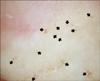
Dermoscopic identification of mites was performed at 10× magnification with a DermLite II Pro hand-held dermoscope (3Gen, San Juan Capistrano, CA, USA). Application of a liquid interface was not required. A triangular structure with following burrow was considered to indicate the presence of a mite (Fig. 1).
Skin scraping
The skin scraping was obtained with a sterile scalpel after the application of 1 drop of mineral oil onto the lesion. Skin scraping was performed two times in each patient. One scraping was observed with dermoscopy (referred to as "with dermoscopy") and the other was performed with the naked eye (referred to as "without it"). Every time a new patient was included in our protocol, the order of the two different scrapings varied so as to prevent any bias in the data resulting from the ordering of the procedure. The number of scraped sites until the examination was declared negative was left to the recognition of the attending dermatologists.
Outcome measures
To compare the efficacy of scraping procedure "with dermoscopy" and "without it", the durations and outcomes of each procedure were measured by timing with a stopwatch. The time was measured beginning from when each patient entered the room for the skin scraping and sat on the chair. Timing was stopped when the doctor declared the skin scraping either negative or positive. To find the specific clinical features influencing difference in the results between the "with dermoscopy" and "without it" conditions, the candidate circumstances suggestively associated with the difference were investigated. The candidate circumstances were the absence of three characteristic symptoms (pruritus with nocturnal exacerbation, visible burrow and contagiousness), previous steroid treatment (history of topical or systemic steroid treatment for more than 2 weeks recently), nodular scabies, infants or the elderly, and immunocompromised host. Finally, we tried to find the specific clinical findings relevant to positive outcomes of "with dermoscopy". The clinical findings included pruritus with nocturnal exacerbation, visible burrow, contagiousness and intensive pruritic nodules on the genital area.
Statistical analyses
The paired t-test and chi-square test were used to investigate if "with dermoscopy" was superior to "without it" in the duration and diagnostic accuracy of the procedure. Additionally, logistic regression was used to analyze the association among the candidate factors and the difference in the result between "with dermoscopy" and "without it". The chi-square test was used again to find possible positive markers. The statistic analyses were carried out using SPSS version 19.0 (SPSS Inc., Chicago, IL, USA). All p-values were two-sided and p<0.05 was considered statistically significant.
RESULTS
Patients
From January 2010 to December 2010, 136 patients were suspected to have scabies and skin scraping with dermoscopy was performed for the diagnosis of scabies. Among them, 117 patients were diagnosed with scabies and 49 patients were enrolled in our protocol.
Diagnostic accuracy of dermoscopy
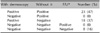
Among 49 patients in whom the skin scrapings were performed, 41 patients were positive "with dermoscopy" and 23 patients were positive "without it". Eight patients were negative in both procedures, and no patient tested was positive for scabies in their next visit. The mean duration of the procedure was 227.1 seconds "with dermoscopy" and 441.9 seconds "without it". In both of the above groups, the results of "with dermoscopy" were statistically superior to those of "without it" (p<0.05) (Table 1, 2).
Clinical circumstances influencing differences in the results between "with dermoscopy" and "without it"
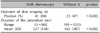
Among 49 patients, 18 showed differences between "with dermoscopy" and "without it". The absence of three characteristic symptoms (nodular scabies, infants or the elderly and immunocompromised host) did not show statistically significant association with the difference between the two. However, previous steroid treatment was associated with a statistically significant association (p<0.05) (Table 3).
Correlation between the presence of specific clinical features and positive outcomes of "with dermoscopy"
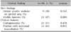
The correlation between the presence of visible burrows and the positive outcomes of "with dermoscopy" was statistically significant (p<0.05), but the other clinical findings were not (Table 4).
DISCUSSION
Scabies is a contagious skin disease that has afflicted mankind for centuries. It is usually transmitted by direct skin-to-skin contact, but fomites play a role in the contagiousness of scabies to some extent8. Epidemiologic studies have linked a higher prevalence of scabies with females and in school-and preschool-age groups, likely reflecting the greater personal contact and socialization among these groups9,10. The most common symptoms are produced by the host immune reaction to burrowed mites and their products. Pruritus and rash may take up to 6~8 weeks to develop after initial exposure to the scabies mite1. For this reason, the early clinical presentation of scabies should be detected as soon as possible to prevent the spread of infestation. Fortunately, scabies does not seem to be highly contagious. A patient with conventional scabies appears to require between 15 and 20 minutes of close contact to transfer the mites to another person2, although it has not been defined clearly.
Ex vivo microscopic examination of skin scrapings performed at appropriate sites is usually the recommended procedure11. However, if the clinical finding of patients with scabies is atypical or the patients are not compliant with the diagnostic procedures, microscopic identification using conventional invasive procedures will not be practical. In these cases, newer methods including epiluminescence microscopy and polymerase chain reaction can be helpful to detect scabies. Among them, 10× magnification examination using a pocket handheld dermoscope can be especially useful. Scabies is common among those in overcrowded conditions, and has been associated with immigrants, poor hygiene, poor nutritional status, homelessness, dementia and sexual contact2. Additionally, patients in these circumstances are also frequently in resource-poor settings. Therefore, portability and ease of use of the pocket handheld dermoscope are very advantageous, especially considering that the diagnosis of scabies is made clinically in this setting6. Also, the dermoscopic examination is painless, which can lead to improved patient compliance, as compared to invasive procedures (Fig. 2).
In this prospective study, we confirmed that the skin scraping "with dermoscopy" is more accurate and faster in diagnosing scabies than those "without it". However, even more surprising was the marked superiority of "with dermoscopy" in diagnosis of scabies compared to "without it". Among 49 patients, 41 were diagnosed by the scraping with dermoscopy, while in contrast only 23 patients were diagnosed without it. The detection rate of "with dermoscopy" was about twice that of "without it". These results mean that we are unable not confirm many of patients with scabies without dermoscopy and the diagnosis could be prone to error. It is highly advisable to use dermoscopy in scraping, considering its low maintenance cost, ease of use and portability, as well as its outstanding performance.
We also tried to find the specific circumstances in which dermoscopic identification is more useful. The study design was based on the premise that dermoscopy is more effective when the patients with scabies present atypical clinical features. Many items such as the absence of three characteristic symptoms, previous steroid treatment, nodular scabies, infants or the elderly and immunocompromised host can cause atypical clinical features of scabies, and so were included as candidate circumstances. Ultimately, this study revealed that a history of previous steroid treatment was associated with the differences in the results between "with dermoscopy" and "without it", and that dermoscopy could be especially helpful in diagnosing patients with scabies incognito. A total of 22 patients in our protocol had been treated with systemic or topical steroids for at least 2 weeks before they visited our clinic. Among them, 21 patients were diagnosed with scabies. We could not find any mites, eggs or feces in 12 of 21 patients without dermoscopy. Also, we experienced several patients with scabies incognito referred to our inpatient department. Some had no typical symptoms or signs of scabies, but were diagnosed with scabies simply by using dermoscopy. Thus, we could prevent spreading of infestation to other patients. The secondary or tertiary care hospital has a better chance to encounter patients with previous steroid treatment than the primary care hospital. The control of the nosocomial infection or infestation could be an important issue in this setting. For these reasons, dermoscopy could be essential in the secondary or tertiary care hospital.
Also, this study revealed the statistically significant correlation between the presence of visible burrows and the positive outcomes of "with dermoscopy". In the case of the absence of dermoscopy or microscopy, for example, the presence of visible burrows in physical examination could be a reliable positive marker in diagnosing scabies.
Although many case reports about experiences of diagnosing scabies with dermoscopy are available, there have been few well-designed studies that have sought to evaluate the diagnostic value of dermoscopy. Because there is no definite criterion for the diagnosis of scabies, it is difficult to design studies to assess the diagnostic accuracy of dermoscopy. Walter et al.6 and Dupuy et al.7 conducted similar studies in which the definition of true-positive, false-positive, true-negative, and false negative findings were based on practical experience with diagnostic tests. While these authors confirmed that dermoscopy itself is a highly sensitive method in diagnosing scabies, the specificity of dermoscopy differed between the studies. The dermoscopic specificity in the study performed by Walter et al.6 (0.46; 95% confidence interval [CI], 0.34~0.58) was much lower than the result of Dupuy et al.7 (0.86; 95% CI, 0.80~0.92). Walter et al.6 also suggested that this was caused by the untrained dermoscopist and patients with pigmented skin. In our opinion, the specificity of dermoscopy is assumed to be higher than that in the study performed by Walter et al.6, but further studies are needed to confirm this hypothesis. Both of the aforementioned studies compared dermoscopy with the skin scraping to assess diagnostic accuracies of the dermoscopy. As has been mentioned, skin scraping "with dermoscopy" is more accurate than the skin scraping itself ("without it"). Therefore, in future studies, the dermoscopy should be compared with the skin scraping "with dermoscopy" instead of the skin scraping itself ("without it") to assess the diagnostic accuracies of dermoscopy.
Considering that dermoscopy has not yet been accepted as a general diagnostic method for scabies and extensive training is required for the observers to avoid confusion of artifacts, the skin scraping with dermoscopy seems to be a choice of method for scabies diagnosis at the present time. In the secondary or tertiary care hospital, we recommend using dermoscopy in diagnosing scabies.


 XML Download