

 PDF
PDF ePub
ePub Citation
 Print
Print


INTRODUCTION
Melanogenesis is a complex, multistage process involving melanin synthesis in melanosomes, the transport of melanosomes to the dendrite tips of the melanocytes, and their release. In melanocytes and in melanoma cells, melanin synthesis is controlled by a cascade of enzymatic reactions. The initial step of melanin synthesis begins with the oxidation of tyrosine to 3,4-dihydroxyphenylalanine (DOPA) by tyrosinase (TYR), the rate-limiting enzyme of melanogenesis1. Melanogenesis is stimulated by various effectors, including paracrine melanogenic factors (α-melanocyte-stimulating hormone [α-MSH], endothelin-1, stem cell factor, basic fibroblast growth factor), cyclic adenosine monophosphate (cAMP)-elevating agents (forskolin, isobutylmethylxanthine, cholera toxin), and ultraviolet B radiation2. Melanogenesis is regulated via various intracellular signaling pathways including protein kinases, such as protein kinase A (PKA)3, protein kinase C-α (PKC-α)4, protein kinase C-β (PKC-β)5, and mitogen-activated protein kinase (MAPK)6,7. The cAMP pathway especially plays a key role in the regulation of melanogenesis through the up-regulation of the key transcription factor microphthalmia-associated transcription factor and subsequent melanogenic enzymes including pre-existing TYR protein and TYR mRNA3,8,9.
Mammalian target of rapamycin (mTOR) is a serine-threonine kinase involved in a number of important cellular processes such as cell growth and proliferation, cell motility, cellular metabolism, and autophagy10,11. mTOR exists within mammalian cells in two functionally and structurally distinct forms of a multi-protein complex, mTORC1 and mTORC2, which take charge of different sets of signaling pathways12. mTORC1 is composed of mTOR, GβL, and raptor, and is rapamycin-sensitive. However, mTORC2, which consists of mTOR, GβL, mSIN1, PRR5, and rictor, is not responsive to rapamycin. mTORC1 can be activated by various stimuli through different upstream molecules in which growth factors affect mTOR via the phophoinositol-3-kinase (PI3K)/Akt pathway13. PI3K initiates a cascade that ultimately results in the activation of Akt. Akt then inhibits the activity of tuberous sclerosis complex 2, which then activates mTOR signaling. S6K1 and 4E-BP1, downstream effectors of mTOR, regulate the translation of specific mRNAs including MITF. Recently, Ohguchi et al.14 reported a negative regulatory role for phospholipase D1 (PLD1) in the melanogenic process through the mTOR/S6K1 pathway in B16 melanoma cells. Furthermore, recent studies have shown that specific inhibitors of PI3K, such as wortmannin and LY294002, stimulate melanin production in mouse and in human melanoma cells, suggesting that the PI3K pathway might be involved in the regulation of melanogenesis15,16. However, direct evidence for the involvement of mTOR in the regulation of melanin synthesis has not been elucidated.
In this study, we investigated whether the PI3K/mTOR pathway is involved in melanogenesis using human MNT-1 melanoma cells. For this purpose, we used rapamycin and wortmannin, potent inhibitors of mTOR and PI3K, respectively. Our results show that rapamycin has a more potent stimulating effect on melanogenesis than α-MSH or wortmannin. Furthermore, combinations of α-MSH with rapamycin or wortmannin show stronger effects on melanogenesis than α-MSH alone. The results are consistent with the conclusion that melanin synthesis is regulated through the PI3K/mTOR pathway, and rapamycin and wortmannin synergize with α-MSH to induce melanogenesis.
MATERIALS AND METHODS
Cell culture
MNT-1 cells are highly pigmented human melanoma cells, and were acquired as gift from Dr. Vincent Hearing (Pigment Cell Biology Section, NIH, USA). These cells were cultured in minimal essential medium supplemented with 20% (v/v) heat-inactivated fetal bovine serum, 10% AIM-V medium, 20 mM HEPES, streptomycin-penicillin (100 µg/ml each), 0.1 mM nonessential amino acids, and 1 mM sodium pyruvate at 37℃ in a humidified atmosphere of 5% CO2 in air. Monolayers of confluent MNT-1 cells were harvested with a mixture of 0.05% trypsin and 0.53 mM EDTA (Gibco BRL, Grand Island, NY, USA).
Cell viability assay
Cell viability was detected using a CellTiter non-radioactive assay kit (Promega, Madison, WI, USA) and estimated by measuring the metabolism of 3-(4, 5-dimethyldiazol-2-yl)-2, 5-diphenyltetrazolium bromide (MTT). Briefly, 300 µl MTT solution (5 mg/ml) was added to each well of a 24-well plate, and cells were maintained for 1 hour at 37℃. Then, 100 µl solubilization solution (50% dimethylformamide and 20% sodium dodecyl sulfate [SDS], pH 4.8) were added to each well. Absorbance was measured at 570 nm, with a microplate reader. Cell viability was expressed as a percentage of cytoprotection versus controls set at 100%.
Measurement of melanin content
Cells were seeded in 6-well plates (Becton Dickinson, Franklin Lakes, NJ, USA) at a density of 1×10⁵ cells per well and were allowed to attach overnight. The cells were then incubated in fresh medium containing various concentrations of rapamycin, dissolved in dimethylsulfoxide at a final concentration of 0.1% v/v. For determination of melanin content, the MNT-1 cells were washed with phosphate-buffered saline (PBS) and dissolved in 250 µl of 1 N NaOH for 1 hour at 80℃ and transferred to 96-well plates. The melanin contents were determined by measuring absorbance at a wavelength of 405 nm.
TYR activity
TYR activity was determined as described previously17 with slight modifications. Briefly, MNT-1 cells were cultured in six-well plates. After incubation with rapamycin for 3 days, the cells were washed in ice-cold PBS and were lysed with phosphate buffer (pH 6.8) containing 1% Triton X-100 and protease inhibitors (Complete™ protease inhibitor mixture; Roche Molecular Diagnostics, Mannheim, Germany). The lysates were clarified by centrifugation for 10 minutes at 10,000 g. After the quantifying protein levels and adjusting concentrations using lysis buffer, 90 µl of each lysate, containing an identical amount of protein, were placed in 96-well plates, and 10 µl 15 mM L-DOPA was added to each well. After incubation at 37℃ for 30 minutes, dopachrome formation was assayed by measuring absorbance at 475 nm using a microplate reader. TYR activity is reported as percentage values.
Western blot analysis
Cells were lysed in cold lysis buffer (0.1 M Tris-HCl, pH 7.2, 1% Nonidet P-40, 0.01% SDS, 1 mM phenylmethylsulfonyl fluoride, 10 µg/ml leupeptin, 1 µg/ml aprotinin) and were centrifuged at 15,000 g for 15 minutes. Total protein contents in the supernatants were measured using a BCA protein assay kit (Pierce, Rockford, IL, USA). Thirty micrograms of protein were resolved by 8% SDS-polyacrylamide gel electrophoresis and were electrophoretically transferred to nitrocellulose membranes, using the semidry technique. After blocking for 1 hour with 5% skim milk in TBS-T buffer solution (10 mM Tris, 150 mM NaCl, and 0.1% Tween-20), each membrane was incubated with primary antibody against TYR, tyrosinase-related protein (TYRP)-1, TYRP-2, MITF, and β-actin (Santa Cruz Biotechnology, Santa Cruz, CA, USA). Specific antibody binding was detected using horseradish peroxidase-conjugated secondary antibody (Santa Cruz Biotechnology), and was visualized by enhanced chemiluminescence detection (Pierce). The amount of protein (arbitrary unit) was quantified using an imaging densitometer and expressed as averages of triplicate experiments.
RESULTS
Effects of rapamycin on melanin content and MNT-1 cell proliferation
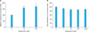
MNT-1 cells were treated with various concentrations of rapamycin for 48 hours. Melanogenesis was then determined by measuring intracellular melanin contents, which are shown as percentage values. Cell proliferation was also expressed as a percentage relative to the control. The effects of rapamycin on melanin content and the proliferation of MNT-1 cells are shown in Fig. 1. Rapamycin showed a melanogenesis-enhancing effect in a concentration-dependent manner (Fig. 1A), but had no significant effect on cell proliferation (Fig. 1B).
Effects of rapamycin on TYR activity in MNT-1 cells
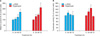
To elucidate the mechanism by which rapamycin stimulates melanogenesis, MNT-1 cells were treated with rapamycin at a concentration of 100 nM. Melanogenesis is regulated by the activity of TYR, the rate limiting enzyme in melanin biosynthesis. Thus, we examined the effect of rapamycin on TYR activity. After rapamycin treatment, melanin contents were significantly increased in a time-dependent manner (Fig. 2A). As shown in Fig. 2B, TYR activity was also increased during the 24~48 hours after the addition of 100 nM rapamycin. According to these results, we hypothesized that the rapamycin-induced melanin formation is mediated via the activation of TYR activity.
Effects of rapamycin on TYR, TYRP-1, TYRP-2, and MITF expression in MNT-1 cells
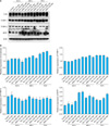
In order to further clarify the mechanism of rapamycin-induced melanogenesis, protein levels of TYR, TYRP-1, TYRP-2, and MITF in MNT-1 cells were determined by Western blot analysis. As shown in Fig. 3A and B, levels of TYR, TYRP-1, and MITF were significantly enhanced after rapamycin treatment in a time-dependent manner compared to the control, whereas there was no effect of rapamycin on TYRP-2 levels. After treatment with 100 nM α-MSH, the protein levels of TYR and TYRP-1 were slightly increased in a time-dependent fashion. These results suggest that rapamycin-induced melanin formation can be mediated via up-regulated levels of TYR, TYRP-1, and MITF proteins.
Stimulatory effects of the combination of α-MSH and rapamycin or wortmannin on melanogenesis
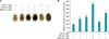
To determine the potential stimulatory effects of rapamycin and wortmannin, specific inhibitors of mTOR and PI3K, respectively, on α-MSH-induced melanogenesis, the cells were treated with α-MSH (100 nM) and/or rapamycin (100 nM) and/or wortmannin (1 µM) for 72 hours. When rapamycin or wortmannin were simultaneously added with α-MSH, melanin synthesis increased dramatically (Fig. 4) in terms of pellet color (Fig. 4A) and melanin content (Fig. 4B). The combination of α-MSH and rapamycin especially showed the most potent enhancing effect on melanin production, eliciting a 3.7-fold increase compared to the 2-fold increase elicited by rapamycin treatment alone.
DISCUSSION
Cell-permeable inhibitors of PI3K/mTOR, such as wortmannin and rapamycin, are potent tools for dissecting intracellular signaling networks. Wortmannin inhibits several protein kinases structurally related to PI3K18 and DNA-dependent protein kinase19, whereas other protein kinases, such as PKA, PKC, and cyclic guanosine monophosphate-dependent protein kinase, are insensitive to wortmannin at concentrations up to 1 µM20. Rapamycin is a potent inhibitor of mTOR which is implicated in regulating cell growth and proliferation12,16.
Melanogenesis is one of the major functions of pigmented cells such as normal melanocytes and malignant melanoma cells. In this study, we demonstrate that the PI3K/mTOR pathway is involved in regulating melanin synthesis, and that α-MSH has a co-stimulatory effect on melanogenesis with inhibitors of this pathway in highly pigmented human MNT-1 melanoma cells. In rapamycin-treated cells, the intracellular melanin content increased in dose-dependent (Fig. 1A) and time-dependent (Fig. 2A) manners. We compared the effects of rapamycin with those of α-MSH, a positive control in those trials because of its known stimulatory effects on melanin synthesis21. Our results show that the stimulatory effect of rapamycin on melanin production in MNT-1 cells was stronger than that of α-MSH (Fig. 2A). To elucidate the mechanism of action of rapamycin on melanogenesis, we measured the enzyme activity of TYR. TYR activity increased significantly at 24~48 hours after the addition of 100 nM rapamycin (Fig. 2B). This enhancing effect of rapamycin was more potent than α-MSH, suggesting that the increased melanogenesis elicited by rapamycin could be achieved, at least in part, by its stimulatory action on the signaling pathways responsible for the regulation of TYR. TYR mRNA levels are generally correlated with TYR activity22. In our study, rapamycin treatment increased the protein levels of MITF, a transcription factor implicated in the regulation of TYR gene expression and melanogenic enzymes such as TYR, TYRP-1, and TYRP-2 (Fig. 3), indicating that MITF up-regulates the expression of TYR and TYRP-1 mRNAs in rapamycin-treated cells. In other words, the rapamycin-induced stimulation of melanogenesis is achieved by the transcriptional regulation of those melanogenic enzymes.
Melanin synthesis in pigment cells is regulated by a complex and interconnected set of intracellular signaling pathways, including PKA (stimulated by cAMP elevators23), PKC (stimulated by endothelins), and MAPK (ERK1 and ERK2; stimulated by growth factors6,7). The cAMP pathway plays a pivotal role in the regulation of melanogenesis through the activation of PKA and cAMP responsive element binding protein (CREB), which leads to upregulation of the expression of MITF3. Among the signaling pathways putatively involved in melanocyte activation, the PI3K/mTOR pathway seems to be a good candidate for studies of pigmentation, since PI3K has been im plicated in the differentiation of dendritic neuronal cells24 and PLD1, an upstream activator of mTOR, exerts a negative regulatory role on melanogenesis through the mTOR pathway in B16 melanoma cells16. Taken together, it is clear that melanogenesis is regulated by a network of several signaling mechanisms including the mTOR pathway. But the precise mechanisms connecting the PI3K/mTOR pathway with melanogenesis have not yet been elucidated.
This study demonstrates that the inhibition of mTOR leads to a stimulation of melanin synthesis that correlates well with increased TYR activity and the protein levels of TYR and MITF. Meanwhile, similar effects were observed with wortmannin, indicating that inhibition of the PI3K/mTOR pathway plays an important role in regulating melanin synthesis. However, wortmannin was markedly less effective in stimulating melanin production than was rapamycin (Fig. 4A). Interestingly, analysis of melanin contents revealed that the combination of α-MSH and rapamycin have a potent co-stimulation of melanin synthesis (Fig. 4B). One possible explanation of this is that the α-MSH-associated cAMP pathway promotes the activation of PKA and CREB, with the inhibition of the mTOR pathway by rapamycin increasing the expression of MITF, thereby simultaneously leading to the induction of melanogenic enzymes such as TYR and TYRP-1.
In summary, we demonstrate that rapamycin, an inhibitor of mTOR, is a positive regulator of melanogenesis in terms of melanin content, TYR activity, and protein levels of melanogenic enzymes as well as MITF. We further show that rapamycin and wortmannin synergize with α-MSH in stimulating melanin synthesis through the inhibition of mTOR and PI3K, respectively. The PI3K/mTOR pathway is involved in regulating melanin production through the regulation of MITF expression. This study performed in melanoma cells has a limitation in terms of pigment cell types. Studies done using normal human melanocytes or in vivo models will be better and more physiologically relevant.
 XML Download