

 PDF
PDF ePub
ePub Citation
 Print
Print


Editor:
In 1896, Fordyce1 described whitish spots on the vermilion border of the lips, oral mucosa and, rarely, genital mucosa. Fordyce's spots are ectopically located sebaceous glands1-4.
To date, few reports have been published to describe the clinicopathologic study of Fordyce's spots in the Korean population. We herein review the clinicopathologic characteristics of Korean patients with Fordyce's spots.
Data were collected retrospectively from the medical records of patients diagnosed with Fordyce's spots between January 2000 and December 2009 in the Department of Dermatology of 4 medical centers in Korea. We reviewed biopsy slides, treatment regimens, and other relevant information. Sixteen patients were finally enrolled in this study. All patients were confirmed with Fordyce's spots following biopsy. This study was approved by the Institutional Review Board of the Catholic University of Korea. Distribution of the patients' age, sex, incubation period, symptoms, signs, anatomical locations, and treatment were evaluated. Histopathologically, the morphology of Fordyce's spots, distribution, and existence of sebaceous ducts-whether they opened onto the skin surface or not-were investigated.
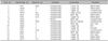
The male to female ratio was 11 : 5. The average patient age was 34.3 years and ranged from 17~67 years (mean±S.D., 34.3±15.0 years). The average age was 41.1 years old in men and 19.4 years old in women. Duration of illness prior to diagnosis ranged from several days to five years, and the mean incubation period was 1.9 years. Most patients were asymptomatic. However, one patient with concurrent contact dermatitis complained of pruritus. The observed lesions were multi-centric and whitish to yellowish in color. Slightly elevated papules and plaques with sizes ranging from 1 to 3 mm were seen. In 8 patients, the lesions were found on the upper lip (50%), on both lips in 4 (25%), and on the lower lip in two (12.5%) (Fig. 1A, B). The spots mostly presented on the vermilion portion of the lips. The lesions in one patient presented on the buccal mucosa (Fig. 1C) and glans penis, respectively. The family history of the patient was unknown. Two patients were treated with a carbon dioxide (CO2) laser, 1 with electrodessication (E/D), 5 with excision of the lesions, and 7 were put on observation. Among those treated, no recurrence was reported (Table 1).
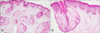
Histologically, the size of sebaceous glands ranged from 1.62±0.55 mm. The glands were located in both the upper dermis (81.3%) and lower dermis (18.7%). The sebaceous glands of Fordyce's spots mostly do not associate with hair follicles. Sebaceous ducts of Fordyce's spots have been observed to open directly onto the surface of the skin (81.3%, Fig. 2).
DISCUSSION
Fordyce's spots are ectopic sebaceous glands on the lips and buccal and genital mucosa (glans penis and labia minor)1-4. The term "Fordyce's condition" has been used on rare occasions5. Although it is infrequent, ectopic sebaceous glands may also be found in the esophagus, gastroesophageal junction, uterine cervix, sole of the foot, thymus, or tongue6-11. A previous study reported a female predilection for Fordyce's spots2. Some studies reported a male predilection or no significant difference in the prevalence between males and females12-14. In this study, they were predominant in men. It has also been previously reported that the incidence of Fordyce's spots increased with age, and 60~80% of patients were elderly15,16. According to Olivier14, Fordyce's spots in a selected South African population reach a peak between 20 and 29 years of age. Our study has shown that most patients were in their early or middle adulthood. We think that the prevalence of younger patients in this study may be due to cosmetic concerns, in addition to a larger proportion of individuals in the younger population who are willing to seek medical help. The vermilion border of the upper lip is the most common site of lesions found on the lip2,12,14. In this study, 16 cases of Fordyce's spots were found on the lips, and one case each on the buccal mucosa and glans penis. In most patients with Fordyce's spots, the upper lip (75%) was involved. There were also several cases in which Fordyce's spots were found on the vermilion border of the lips.
The size of normal sebaceous glands range from 0.2 mm to 2 mm. Ninety percent (90%) of normal sebaceous glands are associated with hair follicles12. The glands may also be found in certain hairless sites, such as Fordyce's spots. Histopathologically, Fordyce's spots are a normal feature of sebaceous glands1-4. They consist of a single sebaceous lobule or gland located in the dermis or submucosa2. The well-formed lobule consists of small clusters of mature sebocytes with a sebaceous duct. This lesion is characterized by the presence of an opening directly onto the epithelial surface. In this study, no hair follicle was observed, and most sebaceous ducts in the dermis open directly onto the surface.
The pathophysiology of Fordyce's spots has not been elucidated. However, since the incidence tends to increase with age, a hypothesis suggests an endocrinary influence on sebaceous glands15. However, this study showed that most cases present in early or middle-aged adulthood. We hypothesize that ectopic sebaceous glands occur with abnormal disposition during embryonic development.
Most patients are asymptomatic; therefore, demand for treatment is not high. However, some patients consider receiving treatment for cosmetic reasons since the lesions do not resolve spontaneously. CO2 laser and oral isotretinoin can be considered as treatment options. However, CO2 laser ablation can leave scars afterwards, and isotretinoin cannot be taken for long periods of time. There is a report on 5-aminolevulinic acid-photodynamic therapy for Fordyce's spots. However, side effects, such as a burning sensation, vesiculation, and post-inflammatory hyperpigmentation have been reported17. Recently, successful therapy that combines CO2 laser ablation and topical trichloracetic acid or bichloracetic acid has been reported, and cauterization with chemical agents is now considered an alternative treatment option for Fordyce's spots. Several patients in this study received treatment for cosmetic reasons and experienced no recurrence after CO2 laser, E/D, or excision.
Sixteen patients who were diagnosed with Fordyce's spots in dermatology clinics from 2000 to 2009 were included in the study. This report is a review upon the first report on Fordyce's spots in Korean individuals, and we expect that this will provide a foundation for further study or medical treatment of patients with this condition.

 XML Download