

 PDF
PDF Citation
 Print
Print


INTRODUCTION
Verrucous carcinoma is an unusual, distinct variant of squamous cell carcinoma, which was first reported by Ackerman in 19481. Verrucous carcinomas consist of oral tumors on the buccal mucosa, which have been referred to as oral florid papillomatosis2. These tumors may be clinically mistaken for giant warts, and both clinical and histological diagnosis must be established. Malignant transformation of verrucous carcinoma into squamous cell carcinoma has been reported in 30~50% of cases3; however, no clear guidelines for effective and safe management of this disease are currently available. Here, we report on two cases of verrucous carcinoma of the lower lip, which were treated with a combination of topical imiquimod (Aldara®) and debulking therapy.
CASE REPORT
Case 1
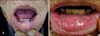
A 70-year-old male patient presented with an extensive verrucous, hyperkeratotic tumor on the central aspect of his lower lip (Fig. 1A). The tumor had been growing for 1 month. The patient was a smoker and a social alcohol drinker. However, his history of trauma to the lesion was unremarkable and he had no past medical history or family history of similar lesions or other skin diseases. Physical examination revealed an exophytic verrucous yellowish-to-whitish tumor measuring approximately 4 cm in diameter. The patient complained of mild tenderness on the lesion and the tumor was firm upon palpation. No lymphadenopathy was detected on the neck or sublingual area.
Microscopic examination of a punch biopsy specimen revealed an endophytic epidermal proliferation with bulbous and blunted projections composed of large well-differentiated squamous epithelial cells extending into the reticular dermis (Fig. 2A). Cellular pleomorphism was minimal and mitotic activity was confined to the basal layer (Fig. 2B). Based on these findings, the patient was diagnosed with verrucous carcinoma. Carbon dioxide laser treatment was then performed following tumescent local anesthesia using lidocaine 0.2%.
After 10 days, the wound was completely epithelialized. At this point, topical treatment with imiquimod was started, with application of cream to the washed and dried tumor surface and then washed off 12 hours after application 3 times per week. After 4 weeks of treatment, the verrucous plaques had decreased in size; however, formation of mild erythema and crust with oozing was observed on the tumor and the patient complained of local pain. Therefore, application of imiquimod was then stopped and the patient received oral administration of analgesics and topical application of antibiotics to the tumor. Over a 4-week period of treatment with analgesics and topical antibiotics, local pain, erythema, and crust slowly disappeared. At this point, administration of imiquimod was started again, with application of treatment 3 times per week over a period of 4 weeks. Treatment was well tolerated by the patient and no unwanted side effects occurred. After 3 months of treatment, the tumor was clinically resolved (Fig. 1B).
Case 2
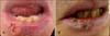
A 75-year-old male developed painful hyperkeratotic plaques on the right aspect of his lower lip, which had persisted for more than 6 months (Fig. 3A). Prior to his visit to our hospital, the patient had been diagnosed with verrucous carcinoma based on the results of a histologic examination conducted at another hospital. Because the lesions progressed in size after the biopsy, he visited our hospital without treatment. The patient was a heavy smoker, but had no previous history of trauma or exposure to radiation.
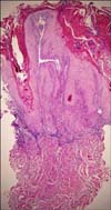
Physical examination revealed verrucous plaques with fissuring on the right aspect of the lower lip. Microscopically, the specimen showed marked parakeratosis, neoplastic proliferation, and downward invasion of epidermal keratinocytes with a blunt border and a pushed-down, bulldozing appearance. Inflammatory cell infiltrates surrounding the tumor were also observed (Fig. 4).
Cryotherapy was performed. Specifically, a cotton stick containing liquid nitrogen was applied to the skin lesion for 2 seconds three times with an interval of five seconds in between each application. Within a few hours of treatment, hyperemia and swelling were obvious and edema and hyperemia increased slowly over a period of 48 hours. The treated mucosa then became covered with a yellowish slough that separated after 7 days. The treated lesion healed completely within 14 days, at which point topical treatment with imiquimod was started, with application of cream 3 times per week. After 3 weeks of treatment, the verrucous plaques almost completely disappeared; however, formation of mild erythema and crust was observed on the tumor, without pain. Topical treatment with imiquimod was continued, with no additional side effects, and, after 7 weeks of treatment, this area was clinically resolved (Fig. 3B).
DISCUSSION
The natural history of verrucous carcinoma of the skin is marked by slow growth of a well-circumscribed tumor, which often shows a papillomatous surface. The tumor is typically located on the sole of the foot or elsewhere on the lower extremities; however, other locations, including the buttocks or the palm of the hand, have been reported4. In 1981, Diaz-Perez et al.5 first reported lip verrucous carcinoma similar to the cases described. In addition, Krutchkoff et al.6 reported that verrucous carcinoma on the lip constituted only 2.8% of all oral neoplasms.
Although the cause of verrucous carcinoma is still unclear, it has been reported to have an association with earlier injuries and scars, as well as with chronic inflammation3,7. Chewing tobacco, betel nuts, and dipped snuffs are also associated with development of verrucous carcinoma of the lip2. Although the role of human papillomaviruses in development of verrucous carcinoma of the skin is still unclear, DNA of human papillomaviruses has been identified in such carcinomas8,9. However, in a clinicopathologic study, Kao et al.4 were unable to demonstrate the presence of virus particles in specimens.
Treatment modalities for verrucous carcinoma include surgical excision, chemotherapy, cryosurgery, intralesional or iontophoretic methods, systemic retinoid therapy, and radiotherapy. Surgical excision is the treatment of choice for verrucous carcinoma. However, incomplete resection can result in accelerated tumor growth; therefore, care must be taken to fully remove all parts of the tumor. In addition, when surgical excision is considered, the size of the defect is often the most important factor because it is usually a strong indicator of the optimal method of reconstruction10. A defect of approximately 30% of the upper or lower lip can be reconstructed by the primary closure due to the great elasticity of the lip11. In our case, total excision was not performed because the tumor encompassed greater than 30% of the lower lip. If a wide excision had been performed in these cases, the defect would have required more complicated reconstructive treatment, such as flap surgery.
Imiquimod, a toll-like receptor 7 agonist, is a patient-applied cream that is used in treatment of certain diseases of the skin, including various forms of skin cancer (basal cell carcinoma, Bowen's disease, superficial squamous cell carcinoma, some superficial malignant melanomas, and actinic keratosis) as well as genital warts11-16. The exact mechanism of imiquimod is not known; however, its effects are thought to be a result of augmentation of immune response through induction of interferon alfa17.
Imiquimod has been used in conjunction with a carbon dioxide laser for treatment of perianal verrucous carcinoma18. In addition, Schalock et al.19 described 2 cases of verrucous carcinoma of the foot, which were treated with imiquimod for more than 1 year.
We did not perform a biopsy after treatment; therefore, we cannot confirm that the tumor has been resolved. However, no further relapse has been observed for 30 months since the last treatment.
In summary, here we describe two cases of rapidly growing oral verrucous carcinoma of the lip, which were treated with a combination of topical imiquimod and debulking therapy. There was no recurrence of the carcinoma for more than 3 months and the cosmesis was excellent. Taken together, these findings suggest that combination therapy of topical imiquimod and a debulking method may be useful for treatment of some cases of verrucous carcinoma.

 XML Download