

 PDF
PDF Citation
 Print
Print


INTRODUCTION
Mycosis fungoides (MF) is a primary cutaneous T-cell lymphoma, which usually starts as "flat patches". Common locations include the lower abdomen, buttocks, upper thighs, and breasts of women1-4. Patches vary in size, with a round, oval, serpiginous, or polycyclic shape, and they have sharp margins1,2. A case of mycosis fungoides on both soles of the feet was recently treated at our center. The lesion was not easily recognized as a flat patch and the diagnosis of MF was delayed. After the correct diagnosis, the plantar lesion required special treatment due to its interdigital involvement and plantar furrows, which were difficult to irradiate with the therapeutic wavelength of electron beam commonly used for this condition3. The patient was successfully treated with three-dimensional conformal radiotherapy (3D-CRT). Here, a case of an 18-year-old Korean woman with MF on the soles of the feet, which was refractory to the usual conventional local treatments, is reported.
CASE REPORT
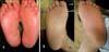
An 18-year-old Korean woman developed hyperkeratotic soles three years prior to presentation to the hospital. For two years before examination, she was treated with oral and topical steroids at local clinics, which caused weight gain and menstrual irregularities, with little improvement of skin lesions. On examination, the soles of both feet showed thick hyperkeratosis with scales and mild erythema. The hyperkeratotic plantar surfaces had shallow furrows. The lesion involved almost the entire plantar surface, including the digital furrows and lateral borders of the soles (Fig. 1). There was no family history of palmoplantar keratoderma.
Biopsy findings of the keratotic plaque were as follows: the epidermis showed compact hyperparakeratosis, acanthosis, spongiosis, and mononuclear cell exocytosis (Fig. 2A). Higher power microscopy revealed intraepidermal collections of mononuclear cells showing hyperchromatic atypical cells that were compatible with Pautrier's microabscesses (Fig. 2B). Infiltrated cells were mostly CD4 positive (Fig. 2C). These findings were consistent with the diagnosis of patchy stage IA of MF.
UVA-1 phototherapy was started immediately with 20 J/cm2 two times weekly (a total of 20 times, total cumulative dose: 400 J/cm2). The disease showed only a slight response. Then, methotrexate 15 mg/week and topical diflucortolone valerate ointment were tried for the next four months, without improvement. Finally, radiation treatment with 3D-CRT using a 6 MV photon beam (Varian, Palo Alto, Ca, USA) was applied to the resistant lesion. To avoid radiation injury to the adjoining joint structures and the surrounding normal tissue, 3D-CRT was applied to plantar lesions and this included interdigital furrows.
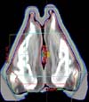
The 3D-CRT procedure was as follows: The patient's legs were placed in a frog leg position. To ensure application of an adequate radiation dose to the skin tissue, a 1 cm-thick silicon bolus was placed on the sole as a tissue compensator. The area for radiation therapy encompassed the normal skin area 2 cm from the margin of the lesion (Fig. 3). A total dose of 40 Gy was administered with a fractionated daily dose of 2 Gy, five days/week for a total of four weeks.
At the time of completion of treatment, the plantar skin had recovered to normal soft smooth skin with mild erythema (Fig. 4A). The post-treatment biopsy examination, performed one month after the end of treatment, showed absence of the previously-present lymphoid infiltrates. All of the lesions were resolved and the patient has been free of disease for one-year of follow up (Fig. 4B).
DISCUSSION
For early stage MF, the goal of therapy is to achieve a complete remission. The choice of treatment method for early stage localized lesions is dependent on the local nature of the lesions or the plaque thickness and the extent of the disease. For early stage MF (stage IA-IIA), Whittaker et al1. recommended that skin-directed therapy (SDT), including topical emollients, steroids, bexarotene gel, UVB/PUVA, and superficial radiotherapy, such as with an electron beam, should be tried as first line therapy.
By nature, plantar skin has a very thick keratinized layer, and an irregular contour, including digital furrows. Therefore, plantar MF is less responsive to skin-directed therapy, compared to other areas of the cutaneous with MF5. Mycosis fungoides and other variants of cutaneous T-cell lymphomas are very radiosensitive; local thick plaque skin lesions can be treated successfully with local radiotherapy6-10.
Technical modifications of local radiotherapy have allowed for optimization of dose distribution, resulting in improved clinical outcome and reduction of chronic complications. The intensity of radiation must be chosen to provide an adequate dose to the bottom of the lesion. An electron beam can be used successfully for treatment of superficial and flat cutaneous lesions of MF; however, for deeper and irregular contoured lesions, electron beam irradiation provides an indistinct dose distribution and ineffective dose delivery to deeper lesions10. A photon beam is usually used for treatment of deep and irregular lesions10. However, for dermatological conditions, this is not commonly used because of the untoward effects on deep regions of the body. Therefore, 3D-CRT using a photon beam was considered for treatment of this patient. The 3D-CRT protocol begins with individualized, 3D digital data sets, which are used in generation of 3D computer images of the patient's lesions and the normal adjacent tissue anatomy. With 3D-CRT, an adequate amount of therapeutic radiation can be delivered to lesions while significantly reducing radiation to the surrounding normal tissues. 3D-CRT increases the effective treatment dose to malignant cells while avoiding harmful effects on normal tissue10. In the present case, a 1 cm-thick silicon bolus was used to provide an adequate radiation dose to the superficial skin layer. A silicon bolus is a tissue equivalent material that is placed directly on the skin surface in order to even out the irregular contours of a patient, which results in presentation of a flat normal surface to the photon beam10.
For successful local radiotherapy, the extent of radiation on the skin is important. Inclusion of the adjoining normal skin, up to 2~3 cm away from the margin of the visible lesions, is recommended, with a total dose of at least 40 Gy9. Otherwise, or with a tight field size without including adjoining normal skin, recurrence at the margins has been commonly reported7. In the present case, radiation treatment with a total dose of 40 Gy, including a 2 cm width of normal skin, was performed.
In this case, satisfactory local control without severe complications was obtained for hyperkeratotic plantar MF. 3D-CRT should be considered for treatment of early stage MF in patients who are refractory to topical agents. However, as local relapse has been commonly reported for many skin-directed therapies1,7, regular follow-up is needed in order to carefully monitor these patients.


 XML Download