

 PDF
PDF Citation
 Print
Print


INTRODUCTION
Papular elastorrhexis (PE) is a rare cutaneous condition characterized clinically by asymptomatic indurated white or flesh papules mainly on the trunk and extremities and histopathologically by decreased and fragmented elastic fibers with or without changes of collagen bundles in the dermis. It commonly occurs during or after adolescence with no history of trauma or local inflammation1,2. Since Bordas et al.1 first used the term PE in 1987, approximately 20 cases have been reported in the literature and the exact origin of this condition remains unknown. We report on a typical case of PE with a literature review focusing on differential diagnosis.
CASE REPORT
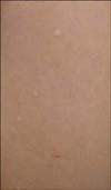
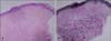
A 12-year-old Korean girl presented with a 9-year history of multiple asymptomatic hypopigmented papules, most of which were several millimeters in size and distributed on her extremities and trunk (Fig. 1). Since its first appearance when the patient was 3 years old, the number of lesions has been slowly increasing. Papules were slightly indurated and scattered with an asymmetric distribution. She had no history of trauma or preceding inflammatory disorders on the involved area. The patient had no family history of similar skin diseases. A biopsy specimen was obtained from a papule on her arm. Histological examination showed hyperkeratotic epidermis and perivascular lymphoid infiltrate in the superficial dermis. Thickness of the layer of finely woven thin meshwork of collagen fibers usually seen in the papillary dermis was observed to increase in the upper dermis with hematoxylin-eosin staining (Fig. 2A). Elastic tissue staining showed primarily decreased and some fragmented elastic fibers in the thickened layer of collagen fibers in the upper dermis (Fig. 2B). Lesions were not treated and persisted during the follow-up period of two years.
DISCUSSION
PE is an uncommon cutaneous condition characterized by multiple asymptomatic papules measuring 1~5 mm, and scattered mainly on the trunk and the proximal portion of the extremities; it occurs predominantly during childhood or adolescence, with a predilection for females3,4. It is also known to have no extracutaneous abnormalities and a lack of family history5. The pivotal histological feature is decrease or fragmentation of dermal elastic fibers with or without changes in collagen bundles in the dermis3,4.
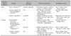
Connective tissue nevi are hamartomas characterized by an imbalance in relative amount and distribution of various dermal connective tissues, including collagen, elastic fiber, or proteoglycan6. PE is regarded as a variant of elastic tissue nevi7,8; however, there is still controversy with regard to whether PE should be categorized as a variant of elastic tissue nevi or as a distinctive entity2,9. Several important disorders require both clinical and histological differentiation from PE (Table 1).
When first reported by Bordas et al.1 in 1987, PE was regarded as a variant of nevus anelasticus due to reduction and fragmentation of elastic tissue. Nevus anelasticus is usually located on mammary areas as nonsymmetrical perifollicular papules and is characterized by prominent loss of elastic fibers rather than fragmentation9. These characteristics help to distinguish it from PE. In addition, in some cases, unlike PE, the disorder has been reported to be congenital8,9. Schirren et al.10 proposed that the disorder may be an abortive form of Buschke-Ollendorff syndrome (BOS) reporting familial PE. On the other hand, Choonhakarn and Jirarattanapochai8 suggested that PE is a unique variant of elastic tissue nevus, not an incomplete form of BOS. BOS is a rare inherited autosomal-dominant syndrome characterized by two different types of connective tissue nevi with or without osteopoikilosis11. Juvenile elastoma is the cardinal elastic lesion showing asymmetrical distribution of yellowish nodules that tend to be grouped into plaques, which is very different from PE; histologically, it shows normal collagen fibrils and an increased number of elastic fibers9,12,13. Dermatofibrosis lenticularis disseminate (DLD), a less common type, shows clinically similar features to PE; however, histopathological study has demonstrated an increase in the number of abnormal collagen fibrils8,9,12,13. We could not find similar skin lesions in the patient's family, which is inconsistent with inheritance of BOS. Ryder and Antaya5 reported that eruptive collagenoma, another variant of connective tissue nevus, showed clinical and histological features similar to those of PE. Recent reports have supported the theory that eruptive collagenoma, nevus anelasticus, and PE represent one disease or disease spectrum1,5,15. No broadly accepted standard for differentiation of this disease from PE has been established; however, the fact that eruptive collagenoma almost always shows prominently increased collagen fibers, while PE may or may not show changes of collagen bundles, may be helpful in distinguishing the two disorders. Due to similar histological changes, familial cutaneous collagenoma (FCC) is another disorder that needs to be ruled out. Although its clinical features also make it hard to differentiate from PE, it clearly differs from PE because it is inherited as an autosomal dominant trait16. Apart from connective tissue nevi mentioned so far, secondary scarring and anetoderma are disorders that should be taken into account when diagnosing PE. Wilson et al.17 reported that distinguishing papular acne scars representing post acne scars from PE was difficult. However, like postacne scars, secondary scarring typically involves follicular papules with a marked decrease of elastic fibers, whereas PE is classically characterized by non-follicular papules with elastorrhexis. Our patient had no history of any preceding inflammatory conditions or trauma on the affected sites and the histological findings showed decreased and fragmented elastic fibers in the upper dermis. Loss of elastic tissue in the dermis is a similar histological feature of anetoderma; however, clinically, it features round, finely wrinkled, atrophic and flaccid patches of skin, which is clearly different from PE18.
 XML Download