

 PDF
PDF Citation
 Print
Print


INTRODUCTION
Localized scleroderma (morphea) usually develops spontaneously. However, a significant number of reports suggest that external stimuli can induce morphea. Although trauma is reported as a morphea trigger, the mechanism has remained unclear. Herein, we report a case of morphea that developed after a needle biopsy and discuss the mechanisms underlying these phenomena.
CASE REPORT
A 35-year-old Japanese female noticed a small breast tumor and underwent a needle biopsy examination. Three months later, she found an egg-sized, slightly itchy erythema on her left breast, around the needle biopsy scar. Subsequently, two walnut-sized erythemas developed. A topical steroid was ineffective. Within a few months, the erythema inflammatory reaction gradually attenuated but sclerotic plaques with slight pigmentation remained. A similar progressing sclerotic plaque had also developed on her right upper arm. She was referred to our hospital with the complaint of these sclerotic plaques.
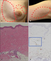
A physical examination revealed an oval sclerotic plaque and two small round sclerotic plaques on her left inner breast (Fig. 1A). An oval sclerotic plaque was observed on her upper right arm (Fig. 1B). No physical findings relevant to systemic sclerosis such as Raynaud's phenomenon, sclerodactyly, or internal organ dysfunction was observed. A histopathological examination of the biopsy specimen from the sclerotic lesion on the upper arm showed a thickened reticular dermis consisting of closely packed, homogenous collagen bundles. A significant number of mononuclear cells had infiltrated the lower dermis (Fig. 1C). Alpha-smooth muscle actin-positive cells had increased significantly in the lower dermis (Fig. 1D). Laboratory data including various auto-antibodies were normal. From these findings, the patient was diagnosed with morphea developing after a needle biopsy.
This patient was successfully treated with oral administration of both suplatast tosilate and tranilast, as well as topical tretinoin tocoferil. Six months later, the first sclerotic lesion on the left inner breast had improved. Other sclerotic lesions on her breast and right arm had completely disappeared.
DISCUSSION
Several studies have suggested that morphea begins with unidentified antigens interacting with the host immune system. Both endogenous and exogenous factors cited previously may influence the disease. Several external stimuli participate in morphea pathogenesis , including trauma or operative stress1, irradiation2,3, infections (viral infections and a Borelia infection4), Bacille Calmette-Guerin vaccination, pregnancy, implantation of a silicon prosthesis, viral factors, toxic factors, and neurogenic factors5. Christianson et al. reviewed 191 patients with localized scleroderma and showed occurrences after trauma (7.3%) and operative procedures (2.6%)6.
Although there is a report that morphea occurred on the site of a penicillin and a local anesthetic injection7,8, in the present case, the morphea developed after a simple physical aseptic needle aspiration without drug infusion. In a previous report, morphea had also developed on the site of an aseptic surgical examination by abdominal laparoscopy in a 56-year-old female patient2. These observations strongly suggest that external physical stimuli can cause morphea. The precise mechanisms underlying these phenomena are unknown. Some speculations include that trauma may trigger the production and release of inflammatory mediators and/or fibrogenic cytokines, such as transforming growth factor-beta, from cells in the microenvironment of the traumatic lesion. This can result in the synthesis of excess collagen in susceptible individuals and lead to sclerosis at a local site9. The reason why sclerosis but not fibrosis is induced with external stimuli and causes morphea in specific patients should be clarified in future studies.
 XML Download