

 PDF
PDF Citation
 Print
Print


INTRODUCTION
Langerhans cell histiocytosis (LCH) is a disease of dysregulated proliferation of Langerhans cells with subsequent organ involvement. Congenital self-healing reticulohistiocytosis (CSHRH) is a congenital variant of LCH, which does not show systemic involvement1. Clinical presentation typically consists of multiple papules or nodules, which show spontaneous involution within a few months. Due to its poorly specific clinical features and self-limiting character, solitary CSHRH is much rarer and difficult to diagnose. Since it is prone to be confused with other dermatologic tumor conditions, dermatologists should have an understanding of the clinical and histologic findings.
CASE REPORT
A 29-day-old girl presented with a solitary skin colored papule with crust on her left sole since birth. She was delivered at term by vaginal delivery following a normal pregnancy to a primipara primigravida mother. Overall, she was healthy, without perinatal problems. There was no Ed-highlight-Please review.
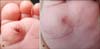
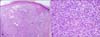
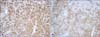
Family history. Physical examination revealed a domeshaped, skin colored papule measuring 7 mm, with crust, on her left sole (Fig. 1A). However, no organomegaly or superficial palpable lymph nodes were observed. Clinical diagnoses, including hemangioma, juvenile xanthogranuloma, and congenital self-healing reticulohistiocytosis were made and skin biopsy was performed on the sole. Atrophic epidermis and effacement of rete ridges were observed (Fig. 2A). A dense infiltrate of histiocytic cells admixed with numerous eosinophils and some scattered lymphocytes were observed. No multinucleated giant cells were observed in our histologic section (Fig. 2B). Histiocytes had a large, vesicular, and some kidney-shaped nuclei with prominent nucleoli. On immunohistiochemical staining with CD1a and S-100, the majority of tumor cells showed strong reactivity (Fig. 3). Ultrastructural studies were not performed. Laboratory studies, including complete blood count, coagulation profile, serum protein electrophoresis, chemical battery, urinalysis, VDRL, congenital infection studies (TORCH), and chest radiography were normal. Two months later, the lesion had completely regressed, leaving a scar (Fig. 1B). At 1 year after diagnosis, no recurrence or systemic involvement has been observed. Based upon clinical, histopathologic, and laboratory findings, a diagnosis of congenital self-healing reticulohistiocytosis was made.
DISCUSSION
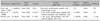
CSHRH, first described by Hashimoto-Pritzker in 1973, is a rare and self-limited form of LCH1. The disease is characterized by multiple papules or nodules, which have a tendency to show spontaneous regression. Lesions are usually multiple, but, more rarely, solitary. The first case of solitary CSHRH was reported by Berger et al.4 in 1986. Since then, several cases of a solitary form have been reported. Bernstein et al.5 reported that solitary CSHRH appeared to contribute to approximately 25% of CSHRH cases. However, this disease shows poorly specific clinical features and spontaneous regression within a few months; therefore, the real incidence of solitary CSHRH might be underreported6. Two cases of solitary CSHRH have been reported in Korea2,3. Reported cases are summarized in Table 1.
Clinical manifestations of CSHRH are polymorphic, presenting as papules, nodules, crusts, vesicles, and, rarely, hemorrhagic bullae, which can sometimes become ulcerated, necrotic, and crusted7-9. Lack of systemic involvement, as well as spontaneous resolution of cutaneous lesions, are essential for a diagnosis of CSHRH. The lesions regress within a mean period of 15 weeks and recurrence has never been reported10. Histopathologically, there are dense intradermal infiltrations of histiocytes showing abundant eosinophilic cytoplasm and kidney-shaped or indented nuclei associated with lymphocytes and eosinophils. These histiocytes show strong expression of S-100 protein and CD1a, markers of Langerhans cells (LC)11,12.
Differential diagnosis includes hemangioma, juvenile xanthogranuloma, Spitz nevi, mastocytoma, and histiocytic disorders. Those diseases can be differentiated by histopathologic findings. In cases of juvenile xanthogranuloma, histiocytes can be differentiated with immunohistochemical staining, such as S-100 protein and CD1a. Among histiocytic disorders, differentiation of indeterminate cell histiocytoma (ICH) from CSHRH is difficult. ICH is characterized by positive immunohistochemical staining for S-100 protein and CD1a, but can be differentiated from CSHRH by absence of Birbeck granules13. Electron microscopic examination of CSHRH reveals Birbeck granules and laminated dense bodies in 10 to 25% of histiocytes14. In our case, we did not perform an electron microscopic examination, but made a diagnosis of CSHRH, since, ICH is much more rare than CSHRH, has tendency to occur in adults, and only a few cases have been reported as a solitary variant7,15.
The pathogenesis of the self-limiting nature of CSHRH remains to be elucidated. Weiss et al.16 have studied various dendritic cell markers in CSHRH. With immunohistochemistry, tumor cells showed positive staining for S-100 protein, CD1a, and HLA-DR. However, tumor cells showed negative staining for Langerin and CD68. They speculated that CSHRH should be comprised of activated mature LCs, because CD68 expression is lost and Langerin staining is decreased in the process of LCs and dendritic cell maturation17,18. The authors explained the self-regressing character of CSHRH as the tumor cells of CSHRH eventually becoming apoptotic on terminal maturation, which is the natural course of LC activation16.
No treatment is required for CSHRH; however, clinical and laboratory monitoring is mandatory. To date, there have been no reports of systemic involvement or complications in solitary CSHRH. However, there has been one case of a girl with CSHRH, who had sparse skin lesions resolving at 1 and 1/2 years of age, but developed diabetes insipidus at the age of 4 years19. Although, there is no evidence of an association of CSHRH with diabetes insipidus in that case, regular follow up will be important for management. Zunino-Goutorbe et al.11 recommended regular physical examination for at least 2 years. They proposed performance of simple laboratory tests and radiographs initially, with repetition of laboratory analyses, including inflammation, hepatic, and differential blood counts at 6 months, and imaging studies only if required by clinical manifestations. We are now following up the patient for one year; however, there is no evidence of systemic involvement and recurrence.
Herein, we report on an additional case of solitary CSHRH with a review of literature.
 XML Download