Abstract
Darier's disease is a genetic disorder of keratinization with autosomal dominant inheritance. Its appearance is usually in the form of greasy, crusted, keratotic yellow-brown papules and plaques found particularly on seborrheic areas of the body. However, there are some clinical variants showing atypical skin lesions. Here we report an unusual case of Darier's disease, which mainly showed prominent comedonal papules over the face.
Darier's disease is a genetic disorder of keratinization with autosomal dominant inheritance, but not always familial1,2. Its occurrence has been found to be due to mutation in the ATP2A2 gene on chromosome 12q23-24, which encodes the sarcoplasmic/endoplasmic reticulum calcium pump ATPase (SERCA2)3-5.
In most cases, it presents with follicular and extrafollicular greasy hyperkeratotic papules and plaques, arising primarily in seborrheic areas6. However, in addition to this hypertrophic subtype, there are other uncommon clinical variants, including vesicobullous, hypopigmented, cornifying, zosteriform or linear, acute, and comedonal subtypes.
Here we report an unusual case of Darier's disease, which mainly showed prominent comedonal papules over the face.
A 31-year-old female presented with asymptomatic multiple skin colored or erythematous greasy papules on the face, which developed in her late 20s. She was otherwise well, without any relevant previous history of skin or medical disease. She said that her relatives on the maternal side, including her grandfather, mother, uncle, two aunts, and two brothers, had the same skin lesions. She had never been treated for her skin problem before.
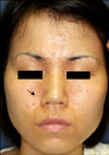
Physical examination revealed multiple skin colored or erythematous papules on the forehead, eyelids, and areas along paranasal and nasolabial folds. Clinically, these papules looked like trichoepithelioma or syringoma, or comedones of acne vulgaris. Her facial skin was greasy (Fig. 1). Otherwise, the scalp, trunk, palms, soles, nails, oral mucosa and teeth were normal.
Skin histopathology of facial papules showed dilated follicular infundibulum, containing keratotic materials with parakeratotic cells. At the lateral aspect and base of the follicular infundibulum, suprabasal acantholysis led to formation of clefts, lacunae, and villi. Dyskeratotic cells, including corp ronds and grains, were observed, as well as brisk perifollicular inflammatory cellular infiltration (Fig. 2).
According to her clinical and histopathological features, we finally diagnosed comedonal Darier's disease. We treated her with oral minocycline and topical tacrolimus ointment; however, her condition showed little improvement and she refused further treatment.
Darier's disease, also known as keratosis follicularis, dyskeratosis follicularis or benign dyskeratosis, is a rare disorder of keratinization that primarily affects the skin. It was described independently by both Darier and White in 1889. It has a prevalence of 1:100,000 of the population and is inherited as an autosomal dominant trait7.
It is characterized by follicular and extrafollicular greasy hyperkeratotic papules and plaques, arising primarily in seborrheic areas6. Vegetating papules, erosions, or hemorrhagic blisters may sometimes be seen. Palms, soles and the oral cavity may be affected. In Darier's disease, there are clinical variants, including hypertrophic, vesicobullous, hypopigmented, cornifying, zosteriform or linear, acute, and comedonal subtypes, similar to our case 2,8-11. Major histopathologic findings include the following: 1) dyskeratosis resulting in formation of corps ronds and grains 2) suprabasal acantholysis, leading to formation of suprabasal cleft or lacunae and 3) villi, which are diagnostic with typical clinical findings.
Previously reported comedonal subtypes presented either as nodular lesions or comedonal lesions, like multiple large blackheads on the face, scalp and the upper trunk. Due to prominent follicular involvement and presence of elongated dermal villi and papillary projections, the histopathological appearance of comedonal Darier's disease differs from that of typical lesions of Darier's disease12. The mechanism of comedone formation in this disease is unclear, but both follicular involvement and dilatation seem to be responsible12. Diseases like acne vulgaris, trichoepithelioma, warty dyskeratoma, familial follicular dyskeratosis and familial dyskeratotic comedone should be differentiated from comedonal Darier's disease. In particular, familial dyskeratotic comedones have very similar features with comedonal Darier's disease, but they present multiple large comedonal papules with a central keratotic plug on the forearms and thighs, whereas the face, scalp and mouth tend to be spared. In addition, corps ronds, lacunae and villi are less prominent in familial dyskeratotic comedones13.
The clinical course of comedonal Darier's disease is often unpredictable and management may be challenging. Treatment with emollients, topical retinoids and topical steroids usually showed limited benefits. Recently, topical tacrolimus has also been tried. Systemic retinoids are often relatively effective, but are toxic. We recommended vitamin A derivatives, but our patient refused it, because she was planning on becoming pregnant. In some uncontrolled studies for flexural and/or hypertrophic involvement, surgical or physically destructive treatments have been used. These include excision, electrodessication, dermabrasion, carbon dioxide, and erbium: ytrium-aluminiumarnet laser abalation6,13.
Comedonal Darier's disease is very rare. Our patient showed scattered multiple papules resembling trichoepithelioma or syringoma on greasy facial skin and, of particular interest, she had a family history of the same skin disease. We could find only six cases of comedonal Darier's disease previously reported in the English literature (Table 1). However, only one of them had a family history. Therefore, herein, we report our interesting case of prominent comedonal Darier's disease with family history, as a very rare one.
Figures and Tables
 | Fig. 1Multiple skin colored or somewhat erythematous papules on the forehead, eyelids and areas along paranasal and nasolabial folds. The facial skin looked greasy (the arrow indicates the biopsy site). |
 | Fig. 2(A) Follicular epidermal invagination, diminished granular layer and lacunae formation. Inflammatory cellular infiltration in the superficial dermis (H&E, ×40 magnification). (B) Keratotic materials with parakeratotic cells in the dilated follicles. Suprabasal lacunae with villi structures (H&E, ×200 magnification). (C) Acantholytic dyskeratosis, corp ronds and grains (H&E, ×400 magnification). |
References
1. Burge SM, Wilkinson JD. Darier-White disease: a review of the clinical features in 163 patients. J Am Acad Dermatol. 1992. 27:40–50.

3. Sakuntabhai A, Burge S, Monk S, Hovnanian A. Spectrum of novel ATP2A2 mutations in patients with Darier's disease. Hum Mol Genet. 1999. 8:1611–1619.

4. Cooper SM, Burge SM. Darier's disease: epidemiology, pathophysiology, and management. Am J Clin Dermatol. 2003. 4:97–105.
5. Jalil AA, Zain RB, van der Waal I. Darier disease: a case report. Br J Oral Maxillofac Surg. 2005. 43:336–338.

6. Aliağaoğlu C, Atasoy M, Anadolu R, Ismail Engin R. Comedonal, cornifying and hypertrophic Darier's disease in the same patient: a Darier combination. J Dermatol. 2006. 33:477–480.

7. Ferris T, Lamey PJ, Rennie JS. Darier's disease: oral features and genetic aspects. Br Dent J. 1990. 168:71–73.

8. Burge S. Darier's disease--the clinical features and pathogenesis. Clin Exp Dermatol. 1994. 19:193–205.

9. Telfer NR, Burge SM, Ryan TJ. Vesiculo-bullous Darier's disease. Br J Dermatol. 1990. 122:831–834.

10. Starink TM, Woerdeman MJ. Unilateral systematized keratosis follicularis. A variant of Darier's disease or an epidermal naevus (acantholytic dyskeratotic epidermal naevus)? Br J Dermatol. 1981. 105:207–214.

12. Lee MW, Choi JH, Sung KJ, Moon KC, Koh JK. Two cases of comedonal Darier's disease. Clin Exp Dermatol. 2002. 27:714–715.

13. Hall JR, Holder W, Knox JM, Knox JM, Verani R. Familial dyskeratotic comedones. A report of three cases and review of the literature. J Am Acad Dermatol. 1987. 17:808–814.


 PDF
PDF Citation
 Print
Print


 XML Download