

 PDF
PDF Citation
 Print
Print


INTRODUCTION
Fixed drug eruption (FDE) is an unusual drug side effect. The disease is defined as recurrent lesions that upon repeated uptake of a causative drug, always appear at the same skin site. The mystery of site preference in FDE is still unresolved. There are a few studies investigating drug-related site involvement in FDE1-3, but there are some anecdotal reports on trauma related localization or on the role of previous inflammation in FDE4,5. Recently, in a Turkish collective of patients, three women with the primary location of FDE lesions on the maximal points of Head's zones were presented6. Their work-up revealed relevant organ disorders with the corresponding projection fields. Head's zones are viscera-cutaneous reflex projection fields on the skin that extend over certain dermatomes and possess a reflex-associated maximal point7,8. This paper describes these 3 cases, and it appears that the primary locations of FDE lie within the reflectorially most active parts (especially buttocks) of viscero-cutaneous referral from a diseased visceral organ (ureter, urinary bladder, kidney, adnexes and uterus), the so-called maximal points of Head's zones.
CASE REPORT
Case 1
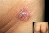
A 68-year-old man presented with an erythematous plaque lesion, 5 cm in diameter, on the skin of the right buttock, which had developed over the previous 3 weeks (Fig. 1). The patient reported monthly reactivation of the lesion, with marked redness and bullae, which healed spontaneously within 5~7 days. Histopathological examination was consistent with the diagnosis of FDE. He had been taking metformin for the last 10 years because of diabetes mellitus. In addition, he occasionally took thiazide for right side ureter stones during 3 months. Upon oral provocation with one-eighth of thiazide, the erythematous lesion reactivated with marked redness and bullae, confirming the diagnosis of thiazid-induced FDE.
Case 2
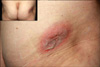
A 79-year-old man presented with an oval shaped, erythematous plaque of 7 cm in diameter on the right buttock area, respectively, following ingestion of a 500 mg tablet of ciprofloxacin (Fig. 2). This was his second episode of development of a patch following ciprofloxacin intake during the last 6 months. He was treated with methylprednisolone, 24 mg/day p.o., for 7 days. Within one week the lesions cleared, leaving residual hyperpigmentation. Histologic examination of a skin biopsy from the skin lesion showed eosinophilic spongiosis and necrotic keratincytes in the basal and upper layers of the epidermis. The diagnosis of FDE was made according to the histology and the history of 2 site-specific attacks definitely following ciprofloxaxin intake. The patient reported right-side chronic pyelonephritis of 2 years, respectively. The location of the lesion on the skin of the patient's right buttock was consistent with the L2-maximal point, the known projection point of the right kidney8,9.
Case 3
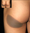
A 23 year-old woman presented with a round shaped hyperpigmented patch, 12 cm in diameter on the left buttock, on exactly the same site as in an episode 3 months previously (Fig. 3). Histopathological examination was consistent with a diagnosis of FDE. Cefotetan and doxycycline were the drugs she had taken occasionally during the last 5 months because of chronic pelvic inflammatory disease. Within 5 days of withdrawal of the suspected drugs, the lesion cleared, leaving residual hyperpigmentation. Three months later, she was treated with doxycyline to control rosacea on her face that led to reactivation of the previous lesion. The case was diagnosed as doxycycline-induced FDE. The location of the lesion on the right buttock area was consistent with the L2-maximal point, the known projection point of the uterus and adnexes8,9.
DISCUSSION
FDE is a common drug side effect constituting about 5~10% of cutaneous drug reactions. The exact pathomehanism of FDE is still unclear. According to recent studies, FDE is a type of IV immune reaction that shows the role of persistent intra-epidermal effector-memory CD8+ T-cells in reactivation, and the role of regulatory CD4+ T cells, transiently migrating into the epidermis of active FDE lesions, in the resolution of FDE10-12.
Clinically, FDE is characterized by the sudden appearance of one or several round, sharply defined erythematous patches and plaque on the skin after drug administration. The lesion has a size of several millimeters up to 10 cm. Sites of predilection are the palms and soles, the medial aspects of the limbs, and abdomen, but also the lips, tongue, and in men the glans penis13. However, little is known about why a FDE lesion comes to its primary manifestation in a certain part of the skin. Studies investigating the predilection areas have indicated 1) drug related, 2) trauma related, or 3) viscerocutaneous reflex related specific site involvement in FDE. It is remarkable that some drugs cause FDE predominantly at specific sites. A highly significant association was found with naproxen and isolated FDE on the lips, and with cortimoxazole and FDE on the glans penis3. On the other hand, some authors have reported the role of a previous trauma in the site preference of FDE4,5,14. Patients were reported in whom FDE lesions initially appeared at exactly the same site as a previous trauma, such as Bacillus Calmette-Guérin vaccination4, burn scars, insect bites, and venepuncture5, or at the site of healed herpes zoster, herpes simplex, or cellulitis14,15. Recently, in a Turkish collective of patients, three women with primary location of FDE lesions on the maximal points of Head's zone were presented6. This work-up revealed relevant organ disorders with the corresponding projection field. The importance of viscerocutaneous reflexes for the location of dermatoses was first recognized in the 1960s7,9. Head's zones are the well known projection areas of visceral organs to the skin via the viscero-cutaneous reflex route16,17. The reflectorially most active parts within these areas were defined by Hansen & Schliack as maximal points of Head's zones7,8. It was claimed that in the case of a visceral pathology, a reflectorially induced optimal alteration in the terrain could arise within the corresponding Head's zones and especially on their maximal points; thus providing a preferential site for a dermatose to come to its primary manifestation. It has been suggested that suitable terrain conditions in the form of a slowing down of blood flow, increased permeability, and the resulting alterations in the metabolism and functions of related skin areas would provide a preferential site for a dermatose to come to its primary manifestation. The first, and up until now, only case of FDE was a patient with an active duodenal ulcer who developed FDE in the duodenal reflex zone of the thoracic dermatomes Th6-Th10 after intake of a pyrazolone derivative9. In the 3 cases reported here, FDE lesions were located on the buttocks consistent with Lumbar 1-3-maximal points, designated as projection points of the kidney, ureter, urinary bladder, adnexes, and uterus to the skin via the viscera-cutaneous reflex route (Table 1). We suggest that any patient with solitary FDE lesions on the buttocks should visit a hospital, and the possibility of visceral organ (ureter, urinary bladder, kidney, adnexes and uterus) pathology should be considered. Although further studies are required to identify the mystery of predilection sites in FDE, viscerocutanoues reflex-related specific site involvement theory may be another optimal model for studying prediction areas in skin disease.

 XML Download