

 PDF
PDF Citation
 Print
Print


INTRODUCTION
Although dermatology textbooks state that sebaceous hyperplasia (SH) is a common normal variant on scrotal skin, many Korean dermatologists consider it rare due to the limited number of reports in Korea. In this report, we describe a patient with SH on the scrotum and penile shaft as a reference case.
CASE REPORT
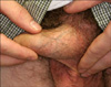
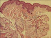
A 26-year-old man was concerned with an increasing number of yellowish papules on his penile shaft without any related symptoms for 2 years. He denied any medical history or manipulation of the site. No family history with a similar disorder was reported. On physical examination, numerous, 1~2 mm-sized papules presented on the proximal half of the penile shaft and parts of the scrotum. The papules were smooth and soft on palpation; a central umbilication was not definite, and many were located around the pubic hair (Fig. 1). We performed a skin biopsy from a representative yellow papule. A microscopic examination showed a lobulated lesion composed of enlarged sebaceous glands (>4 around each pilosebaceous unit) extending down into the dermis. The cells were predominantly mature sebocytes with a peripheral germinative layer. The cells displayed a foamy, vesiculated cytoplasm and a central nucleolus with no atypical features (Fig. 2). Serial sections further showed that the sebaceous lobules were connected with a central pilosebaceous follicle. Based on this evidence, a diagnosis of SH was made.
DISCUSSION
Sebaceous glands form part of the skin appendages. They present in large numbers on the face, scalp, and ears in association with hair structures1-4. Free sebaceous glands (those not associated with the hair follicle) are occasionally found in some areas of modified skin, such as the nipple (Montgomery tubercles), lips (Fordyce's spots), and the inner surface of the prepuce (Tyson's glands)5.
Facial SH in the elderly is due to decreased cellular turnover secondary to reduced androgen levels, with a resulting increase in gland size6. However, this does not explain the hyperplasia that occur in the glands of the penile shaft. In contrast to facial lesions, the majority of penile SH cases seem to appear at an earlier age. Higher sensitivity of the sebaceous cells to androgens, leading to an increase in cellular proliferation may be a possible cause of penile SH7. Inflammatory stimulation by CD8+ lymphocytes also plays a role8.
The clinical differential diagnosis includes milium, lichen nitidus, molluscum contagiosum, genital warts, ectopic sebaceous glands, and pearly penile papules. Dermoscopy and a histological examination can aid in the diagnosis of penile SH. Dermoscopy is a simple and non-invasive tool, which is widely used in the dermatologic field. SH is viewed as aggregated white or yellow nodules with small craters and "crown vessels" (non-arborizing vessels) under dermoscopy8.
SH has been described in textbooks as a common normal variant on the scrotal skin and penile shaft9, but it has rarely been reported in Asia. We do not know the true incidence of scrotal SH in Asians, but it is very likely that many Koreans are reluctant to discuss skin lesions on their genitalia, which makes its overall incidence appear low. Accordingly, only one report has been published on SH of the scrotum and penile shaft in Korea. In Choi's study10, the authors reported their case as unusual, which may not be true. We feel that our case would be an additional good normal variant reference, particularly for Asian dermatologists.
SH should be considered a possibility when facing a patient with multiple tiny papules on the penis. It is important to reassure the patient that SH is not contagious. Treatment is not necessary other than for cosmetic purposes.
 XML Download