

 PDF
PDF Citation
 Print
Print


INTRODUCTION
Ulcerative colitis (UC) is a subgroup of inflammatory bowel disease (IBD). It is diffusive inflammation limited to the large intestinal mucosa. There can be many extragastrointestinal symptoms with the progress of IBD. Skin manifestations such as pyoderma gangrenosum and erythema nodosum usually occur in conjunction with Crohn's disease and UC. However, other skin diseases, such as leukocytoclastic vasculitis (LV) has rarely been reported. Skin manifestations are important clues for diagnosis because they may precede gastrointestinal symptoms1.
LV is characterized by neutrophilic invasion and fibrinoid necrosis along with endothelial enlargement in postcapillary venules. It is a syndrome in which patients most commonly present with palpable purpura on lower extremities and ankles2. An annular variant of LV is rare and to date, only 15 cases have been reported3.
Association between annular leukocytoclastic vasculitis (ALV) and UC is very uncommon and to the best of our knowledge, this is only the second report. We report a case diagnosed as ALV 5 years after the development of UC, which is highly rare.
CASE REPORT
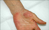
A 66-year-old woman presented with painful polycyclic erythema on both palms. She had been diagnosed with UC 5 years earlier. Her UC had been well controlled with mesalazine and aspirin since June 2006. She was hospitalized in April 2009 due to gastrointestinal tract bloody excrement caused by worsening of the UC. The dose of mesalazine was increased and a fair response to the symptoms of the UC was observed. In June 2009, 4 days before visiting our department, confluent annular erythema appeared on both palms. The pain was not severe, but tenderness was accompanied and the lesions became larger. Therefore, she was referred to our department. Her family history was unremarkable and there was no previous history of any skin disease or skin allergy.
The laboratory test showed elevated erythrocyte sedimentation rate: 66 mm/hr (normal: 0~20) and elevated C-reactive protein: 3.48 mg/dl (normal: 0~0.05). The complete blood count and routine chemistry were within normal range.
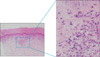
Multiple confluent annular erythematous patches were observed on both palms (Fig. 1). A skin biopsy performed on an erythematous part of the palm demonstrated the presence of lymphohistocytic infiltration around the blood vessels of the upper dermis, edema of endothelial cells and extravasation of erythrocytes (Fig. 2). Diagnosis of ALV was confirmed. The patient was treated with dapsone 200 mg/day, aceclofenac 200 mg/day, levocetirizine 5 mg/day and fexofenadine 180 mg/day for 1 week following biopsy. Her symptoms and signs were completely gone without sequelae.
DISCUSSION
Extra-intestinal manifestations of IBDs including mucocutaneous involvement and skin manifestations occur in about 15% of patients with inflammatory bowel disorders4. The most common skin manifestations are pyoderma gangrenosum and erythema nodosum, while necrotizing vasculitis, cutaneous polyarteritis nodosa and granulomatous perivasculitis are less frequently observed5. In the present case, intestinal manifestations of UC were associated with uncommon skin lesions characterized by confluent annular erythema, which was diagnosed as ALV. Since the skin lesions developed 1~2 months after the UC worsened, we speculated that there may be an association between the two conditions.
ALV is not a clear status within LV, classified based on the clinical morphology. Cribier et al. first reported that some patients with ALV constitute a distinct subtype, which satisfies the following criteria6. The characteristics are: (1) multiple attacks over years with sudden onset and spontaneous regression after 7~10 days, (2) annular purpuric patches that show centrifugal extension, (3) extension over the limbs and trunk creating polycyclic lesions that clear leaving mild haemosiderin deposition, (4) no extracutaneous symptoms and good general health, (5) histological changes of LV with mild vascular changes and intense polymorphonuclear cell infiltration, and (6) complete clearance of all lesions with dapsone6. Our case matches the second, fifth and sixth criteria established by Cribier. Therefore, we cannot classify this case as a "distinct subtype"; however, we thought that there was enough evidence to categorize this case morphologically as ALV. Also, the most potent treatment for ALV is dapsone.
There have been about 20 cases in the literature describing ALV in association with systemic diseases such as monoclonal gammopathy, sarcoidosis, UC, myelomatosis, pregnancy and chronic hepatitis B 3,7-10. Association between ALV and UC is very uncommon and, to the best of our knowledge, this is only the second report.
The etiology of LV with UC is not clear; a partial defect of immunity common to the skin and intestines has been suggested11. The causes of immune activation are believed to be due to the presence of antigens related to infectious agents, drugs, neoplasia, food, or autoantigens12. Attracted neutrophils due to immune activation release lysosomal enzymes, which induce nitric oxide (NO). NO causes apoptosis of neutrophils leading to leukocytoclasis13. At present, the clinical characteristics of ALV, annularity and peripheral spreading, are not known. However, it is possible that inflammatory cells, inflammatory mediators and antigens spread peripherally resulting in an annular pattern of erythema14.
One possible hypothesis for the association between ALV and UC is that the pathogenesis of both is based on immune mechanisms and deposition of immune complexes in the vascular structure and intestinal mucosa for ALV and UC, respectively15.
Skin lesions of LV can be treated with corticosteroids, dapsone, colchicine, or immunosuppressive agents2. In contrast, in ALV, the most potent and effective treatment is dapsone at a daily dosage of 100 mg to 200 mg.
Although ALV in patients with UC is a very rare combination, clinicians need to be aware of this possible association and the most effective treatment for ALV is dapsone.
 XML Download