

 PDF
PDF Citation
 Print
Print


INTRODUCTION
H1 antihistamines are widely used and probably most frequently used in allergic diseases. Topical application of antihistamines commonly leads to sensitization for patients; however, skin reactions provoked by systemic administration of antihistamines have been very rare1,2. Although the rate of urticaria induced by antihistamines has been rare, its relevance should not be ignored. To our knowledge, only 16 cases of H1 antihistamine-induced urticaria have been reported in the literature2-13, and it has never been reported in Korea. Herein, we report a rare case of fexofenadine-induced urticaria.
CASE REPORT
A 69-year-old man first visited at our dermatology department with a 10 month history of pruritic papules on his face and scalp in January 2009. With the diagnosis of allergic contact dermatitis, he had been receiving treatment with levocetirizine (Xyzal®) the previous week with no improvement. During the first three days of fexofenadine (Allegra 180®) supplementation, he experienced episodes of itchy hives over his body about two hours after receiving 180 mg of fexofenadine in addition to 5 mg of levocetirizine. After ceasing the fexofenadine for two days, the symptoms disappeared; however, it recurred after rechallenging with the same drugs the next day.
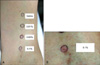
Personal and family histories were not remarkable. Dermatological examination revealed pruritic wheals over his body. He was treated with oral corticosteroids for three days and subsequently, levocetirizine was continued to control allergic contact dermatitis. No further wheals were observed. Three months later, prick tests with fexofenadine dilutions of 0.01%, 0.05% and 0.1% (powder dissolved in 0.9% NaCl) were performed. After ten minutes, a wheal developed at the 0.1% fexofenadine site (Fig. 1). Patch tests with various classes of antihistamines including fexofenadine, terfenadine, and levocetirizine were carried out; however, but the results were all negative (powdered in petrolatum with concentrations of 1%, 5%, and 10%, data are not shown).
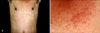
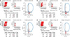
To confirm the diagnosis, oral provocation tests were performed. Increasing doses were administered at 1-hour intervals starting with the half-dose. Urticarial eruptions developed over the body one hour after taking the total dose (180 mg) of fexofenadine (Fig. 2). Flow cytometryassisted basophil activation test with the patient's blood showed positive results (Fig. 3).
DISCUSSION
Fexofenadine is an active metabolite of terfenadine, and is a second generation antihistamine derived from piperidines. Adverse reactions to antihistamines have been rare, and second generation antihistamines are also more selective and less sedative. The most common adverse reactions related to fexofenadine have been headache, dizziness, daytime drowsiness, nausea, among others at a rate of more than 1%, and it has been very rarely reported to cause hypersensitivity14. Cutaneous lesions have been demonstrated to appear as urticaria; maculo-papular, morbilliform, and scarlatiniform eruptions; erythema multiforme; photosensitivity; fixed eruptions; and anaphylactic shock1.
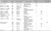
Sixteen cases of antihistamine-induced skin reaction in the form of urticaria have been reported in the literature (Table 1), with cetirizine being the most common causal drug, and diphenhydramine, prophenpyridamine, loratadine, mequitazine, levocetirizine, ebastine, bepotastine, hydroxyzine, olopatazine and dexchlorpheniramine also have been reported to cause urticarial reactions. Fexofenadine has been identified as the eliciting drug in three previous cases6,11,12.
In most cases of antihistamine-induced urticaria, oral provocation test and prick tests were performed. Oral provocation test results were positive in all cases; however, prick test results were positive in only five cases including our own2,4,9,13.
The mechanism of antihistamine-induced urticaria has remained controversial. In some reported cases, it has been noted as a type I IgE-mediated hypersensitivity reaction because of a suggestive clinical history and positive reaction on prick and intradermal tests4,12,13. In contrast, other reports have shown negative results to prick tests with the causative drugs, suggesting a non-immunologic reaction. Possible mechanisms include a Type IV hypersensitivity reaction, non-specific mast cell degranulation or activation of the alternative pathway of the complement system7, or an intolerance reaction9,10.
The basophil activation test is a functional in vitro test that has been used to investigate the cause of allergic reactions. Basophils are now considered equivalent to tissue mast cells since they play, by themselves, a pivotal role in the immediate allergic reaction. Specific population of cells can be identified by flow cytometry, which is a reliable tool for monitoring basophil activation upon allergen challenge by detecting surface expression of degranulation/activation markers, most commonly CD63 and CD203c. The flow cytometry-assisted basophil activation test has become a standard tool for in vitro diagnosis of immediate allergy15.
In our patient, a commercialized Flow-CAST kit® (Buehlmann laboratories AG, Schoenenbuch, Switzerland) was used with fexofenadine at 1.25 mg, 2.5 mg and with stimulation buffer and anti-IgE receptor antibody acting as negative and positive controls, respectively. Peripheral blood leukocytes were isolated from patient's whole blood samples and primed with stimulation buffer containing interleukin 3. Fexofenadine 1.25 mg and 2.5 mg were added and the cells were incubated to mimic the in vivo situation where specific IgE bind to the cellular surface, are bridged by the allergen, and activate intracellular enzymatic cascades leading to the activation of basophils. During the activation, intracellular compounds containing the transmembrane protein CD63 fuse to the cellular membrane and are expressed to the extracellular matrix. Activated basophil levels of fexofenadine-stimulated samples were 44.73% and 10.58% (fexofenadine 2.5 mg and 1.25 mg respectively) in our patient, which was higher than the negative control (4.57) and therefore, the test was positive to fexofenadine (Fig. 3).
Herein, we report an interesting case of urticarial reaction due to fexofenadine. The precise mechanism of urticaria induced by antihistamines has not yet been elucidated. In our case, urticarial eruptions developed one hour after taking fexofenadine prick test, oral provocation test with fexofenadine, and the basophil activation test showed positive results and thus, an underlying immune mechanism cannot be ruled out. Although the allergy tests could not distinguish the precise mechanism of the reactions, they did demonstrate the causal drugs and allowed for the discovery of a well-tolerated drug of the same class. Physicians must be aware that, occasionally, drugs used in treatment act as the causal agent themselves.
 XML Download