

 PDF
PDF Citation
 Print
Print


INTRODUCTION
Acrodermatitis enteropathica (AE) is a rare hereditary or acquired disorder of zinc deficiency first described in 19421. The condition is characterized by alopecia, diarrhea, lethargy, acute eczematous and erosive acro-orificial dermatitis, and acute paronychia2. A rare variant of AE, bullous AE, has unique histopathological findings encompassing clustered and individual necrotic keratinocytes, intraepidermal vesiculation with scant spongiosis, and a mostly lymphoneutrophilic infiltration within the vesicles3. We experienced a case of bullous AE secondary to total parenteral nutrition (TPN) for the treatment of acute pancreatitis occurring in a six-year-old male with acute lymphocytic leukemia who underwent chemotherapy. To our knowledge, this is the first report of bullous AE in the Korean dermatologic literature. Here, we report our case with a review of the associated literature.
CASE REPORT
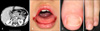
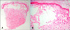
A six-year-old male was transferred to the Catholic Hematopoietic Stem Cell Transplantation Center in December, 2008 with acute pancreatitis, hypertension, and seizure secondary to chemotherapy initiated one month previous for acute lymphocytic leukemia. The chemotherapy regimen consisted of prednisone, vincristine, daunorubicin, L-asparaginase, and methotrexate. Abdominal computed tomography performed prior to transfer to our facility showed diffuse edematous swelling of the pancreas and extensive peripancreatic fluid collection (Fig. 1A). Brain magnetic resonance imaging revealed bilateral asymmetric high-signal intensity in both temporo-occipital and parieto-occipital lobes with some cytotoxic edema in the right occipital lobe. Laboratory investigations revealed a white blood cell count of 1,520/mm3, total bilirubin of 3.1 mg/dl (normal range, 0.2~1.0 mg/dl), direct bilirubin of 0.8 mg/dl (normal range, 0~0.5 mg/dl), amylase of 919 IU/L (normal range, 37~150 IU/L) and lipase of 2,559 U/L (normal range, 114~286 U/L). Acute pancreatitis had developed as the result of L-asparaginase complication and posterior reversible encephalopathy syndrome due to acute hypertensive encephalopathy or chemotherapeutic agents. Amlodipine was prescribed to control the hypertension. Chemotherapy was stopped and TPN was started to treat the acute pancreatitis. After one month, the patient was referred to the dermatology department for consultation regarding periorificial, reddish, eroded bullae with multiple vesicles and blisters on his fingers, toes, and buttock that had developed five days previously (Fig. 1B~D). Bullous disorders such as chronic bullous disease of childhood, herpetic whitlow, and hand-foot-mouth disease were suspected. A Tzanck test of the bullae was negative. A biopsy was subsequently performed of the bulla on the right second finger. Histopathology revealed necrotic keratinocytes with multiple intraepidermal vesicles and perivascular infiltration with predominant lymphocytes and few neutrophils within the dermis (Fig. 2). Based on the histopathological findings, bullous AE was suspected. The diagnosis was confirmed by chemistry results showing reduced serum zinc of 16.5 µg/dl (normal range, 66~110 µg/dl) and alkaline phosphatase (ALP) of 35 IU/L (normal range, 80~220 IU/L). Skin lesions improved rapidly within a few days of starting intravenous zinc sulfate supplementation.
DISCUSSION
AE cases secondary to TPN have been reported previously4,5. TPN solutions contain glucose, amino acids, and principal electrolytes including Na, K, Cl, Ca, and Mg without vitamins and trace elements4. Therefore, in about two weeks, zinc levels decrease below the normal range and symptoms develop6. Zinc functions in the formation and maintenance of all tissues7. The skin includes approximately 6% of total body zinc8. In addition, zinc is an essential component of various metalloenzymes such as ALP, carbonic anhydrase, and RNA polymerase7. Of those elements, ALP is a readily available test that it worthwhile to perform7. In this case the decrease in ALP was indicative of its diagnostic value.
Keratinosomes are enriched in metalloenzymes and they control keratinization9. Therefore, zinc deficiency affects normal keratinization and gives rise to keratinocyte degeneration10. Histopathologically, bullous AE is accompanied by highly eosinophilic, necrotic keratinocytes, and intracellular edema bringing about intraepidermal vesiculation3,11. These histopathologic features of bullous AE distinguish it from spongiotic dermatitis. Cases of bullous AE show prominent vesiculation in the midst of absent to scant spongiosis, adjacent eosinophilic to necrotic keratinocytes, and primarily lymphoneutrophilic infiltration3. Histopathological findings in our case were consistent with bullous AE.
Recently, atypical histopathologic findings in a patient with Crohn's disease who presented with bullous AE were reported12. Skin biopsies revealed intraepidermal vesiculation with eosinophilic necrotic keratinocytes, marked vacuolar degeneration of basal keratinocytes, and lichenoid interface dermatitis. As a result, the authors initially considered lichenoid drug eruption with bullae formation; but the worsening skin lesions upon TPN shifted suspicion to zinc deficiency. Therefore the authors suggested that clinical suspicion of AE is more important than histopathologic findings in diagnosing AE.
Treatment for AE entails 3 mg/kg/day of elemental zinc supplement. Once replacement therapy is initiated, clinical manifestations improve within days to weeks7. In our case, there were multifactorial issues related to zinc deficiency including inadequate intake of zinc related to TPN, malabsorption caused by acute pancreatitis and methotrexate13, and increased metabolism due to leukemia. In conclusion, when using TPN, special attention should be paid to prevent zinc deficiency. Owing to the rarity of the disease, if clinicians do not initially suspect the bullous form of AE, diagnosis and treatment will be delayed; therefore, clinicians must consider bullous AE when they encounter bullous dermatosis. Skin biopsy is an essential component of the workup necessary to diagnose bullous AE.
 XML Download