

 PDF
PDF Citation
 Print
Print


INTRODUCTION
Porokeratotic eccrine ostial and dermal duct nevus (PEODDN) is a rare cutaneous disorder considered to be an eccrine hamartoma, which was first described by Abell and Read in 19801. It is characterized by multiple keratotic papules or plaques and punctate pits with a linear distribution usually located on the extremities along Blaschko's lines. PEODDN is a congenital dermatosis with a tendency to persist, and it usually grows as the patient grows up2. There is no definitive treatment for PEODDN to date, and several treatment methods show unsatisfactory results in regard to therapeutic effects3. We describe a case of PEODDN showing partial remission after 2 sessions of topical photodynamic therapy (PDT). To the best of our knowledge, PEODDN treated with PDT has not been previously reported in the literature.
CASE REPORT
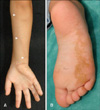
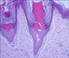
A 4-year-old girl was presented to our hospital with asymptomatic skin lesions on her right upper and lower extremities that had been present since birth. Physical examination revealed multiple keratotic papules on the flexural side of the right ring and little fingers, punctate pits with central keratotic plugs on the right palm and sole, and verrucous plaques on the right forearm in a linear distribution along Blaschko's lines (Fig. 1, Fig. 2A). She had no personal history of extracutaneous abnormalities and no family history of similar lesions. A 3-mm punch biopsy was performed at the plaque on the right forearm. Histopathological examination demonstrated cornoid lamella-like parakeratotic columns connected with dilated acrosyringium within epidermal invaginations, absence of the granular layer, and numerous dilated dermal eccrine ducts. Adjacent epidermal cells showed vacuolization. In the upper layer of the dermis, a minimal lymphocytic infiltration was present around blood vessels (Fig. 3). She was diagnosed as PEODDN and treated with 0.05% tretinoin cream for 3 months and received 2 sessions of CO2 laser ablation for the lesions on the right hand, but the lesions failed to improve.
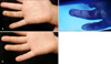
Therefore, we decided to try topical PDT on the same refractory lesions. The superficial scales were removed by a surgical blade and 20% 5-aminolevulanic acid (ALA, Levulan®, MEDEC, Hamburg, Germany) was applied on the right finger lesions under an occlusive dressing for 4 hours. The uptake of 5-ALA to the lesions and peri-lesions was identified with fluorescence photography under Wood's light (Fig. 2B). After injecting local lidocaine to reduce pain, irradiation with red light 633±6 nm LED device (Omnilux revive™, Montgomeryville, PA, USA) at a fluence of 126 J/cm2 was applied to the patient. The ALA-PDT treatment was repeated every 2 weeks for a total of four treatments (right finger lesions, on first and third treatments; right palm lesions, second and fourth treatments). After 2 PDT sessions, additional treatment was not performed because the patient and her mother refused further treatment due to PDT-associated pain. One year after the last treatment, skin lesions remained reduced in size or number (Fig. 2C).
DISCUSSION
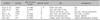
PEODDN is a rare benign cutaneous disorder considered to be an eccrine hamartoma, and only 5 cases of PEODDN have been reported in the Korean dermatological literature (Table 1)4-8. PEODDN is clinically characterized by punctate hyperkeratotic papules, which generally show a linear distribution on the extremities9. Palms and soles are the most commonly affected sites, but PEODDN may occur at other areas of the extremities and even on multiple locations. A verrucous component can be noted when lesions spread out from palmar or plantar surfaces to other sites of the extremities3. Histopathologically, PEODDN is characterized by cornoid lamellae connected with dilated eccrine acrosyringia within an epidermal invagination, which is pathognomonic9. Vacuolated keratinocytes can also be seen within epidermal invaginations10. The differential diagnoses of PEODDN consist of several disorders showing similar clinical findings, including nevus comedonicus, linear verrucous epidermal nevus, and punctate porokeratosis5. Nevus comedonicus and linear verrucous epidermal nevus can be differentiated from PEODDN by the absence of punctate pits and keratin plugs3. Punctate porokeratosis is usually observed in adults but is also observed in congenital forms5. Congenital unilateral punctate porokeratosis may clinically resemble PEODDN because it shows multiple punctate pits and keratotic plugs on the extremities. However, in punctate porokeratosis, no eccrine duct hyperplasia is present, and the epidermal invagination is thinner than that of PEODDN11.
The histogenesis of PEODDN is unclear. While some investigators have suggested that the epidermal invagination of PEODDN originates from a widely dilated acrosyringeal duct that is contiguous with the dermal duct at its base12, others have suggested that the epidermal invagination represents an abnormal clone of epidermal cells that produce a cornoid lamella-like column13. Masferrer et al.3 suggested PEODDN originates from a circumscribed keratinization abnormality pertaining to the hyperplastic and abnormal acrosyringia based on positive carcinoembryonic antigen staining along the ductal lamina through the parakeratotic column of the cornoid lamellae. PEODDN is a persistent lesion and may become enlarged as the patient grows up. Treatment is directed at minimizing cosmetic disfigurement. Various therapeutic modalities have been proposed to treat PEODDN, including topical steroids, topical calcipotriol, retinoid, cryotherapy, and carbon dioxide laser ablation; however, they usually have unsatisfactory results except for surgical excision. We tried carbon dioxide laser ablation in this case, but we had difficulty treating the patient because of the pain. In addition, there is a possibility of leaving a scar when ablating deep into the dermis. Therefore, we applied the carbon dioxide laser at a low energy density, which could be responsible for the unsuccessful outcome of the treatment.
The best treatment strategy for PEODDN is to reduce lesions with little damage to normal peri-lesional skin2. As applying topical fluorouracil does not cause too much pain, it might be beneficial for the patient. Yet no topical agent consisting solely of fluorouracil is licensed in the current Korean pharmaceutical market. As a result, we tried to treat the patient with topical PDT. The mechanism of PDT is based on the interaction of three components: photosensitizer, light, and oxygen. PDT uses light to activate a photosensitizer in lesional tissue, cascading to the formation of cytotoxic reactive oxygen species and selective tissue damage. Generally, PDT is mainly used to treat premalignant lesions, such as actinic keratosis and Bowen's disease, in addition to superficial nonmelanoma cancers14. It was reported that porokeratosis of Mibelli was successfully treated with combined use of 5-ALA, PDT, and fluorouracil. Porokeratosis is formed by a clonal hyperproliferation of atypical keratinocytes, resulting in the cornoid lamellae. Because PDT selectively targets active, atypical cells and causes destruction by the creation of toxic intermediates, it appears to be effective for treatment of porokeratosis15. As atypical hyperproliferation of keratinocytes and cornoid lamellae are common features of PEODDN and porokeratosis, we assumed PDT to be also effective for treatment of PEODDN and therefore decided to apply PDT to this case.
In our case, 5-ALA PDT showed partial efficacy after 2 sessions of treatment. To our knowledge, although it showed partial response, this is the first report of PEODDN treated with topical PDT. Further follow-up and treatment are needed to confirm whether additional sessions would be needed to acquire complete remission. In addition, conducting controlled studies with long-term follow-up would be worthwhile in patients with PEODDN to evaluate its real effectiveness and to compare different treatment modalities for PEODDN.
 XML Download